



Beyond Valve Replacement: Why Procedural Success Does Not Equal Cardiac Recovery
Transcatheter aortic valve replacement has fundamentally changed the therapeutic landscape of severe aortic stenosis. By relieving the fixed obstruction at the aortic valve, TAVR rapidly reduces left ventricular outflow resistance and can interrupt the chronic pressure-overload state responsible for ventricular hypertrophy, elevated myocardial wall stress, diastolic dysfunction, and eventually systolic impairment. In many patients, this unloading is followed by left ventricular reverse remodeling, improvement in ejection fraction, regression of hypertrophy, reduction in filling pressures, and improvement in symptoms. Yet an important clinical paradox remains: a technically successful TAVR does not guarantee recovery of the myocardium or resolution of heart failure.
This distinction is increasingly important as TAVR is performed across broader risk groups and in patients with increasingly complex cardiovascular phenotypes. Procedural success is generally judged by appropriate prosthetic valve deployment, acceptable transvalvular gradients, absence of major complications, and satisfactory vascular and conduction outcomes. These endpoints are essential, but they describe the intervention rather than the biological response of the patient. A valve can function normally while the ventricle remains severely remodeled, fibrotic, ischemic, stiff, or mechanically inefficient.
Severe aortic stenosis produces a prolonged pressure-overload state. Initially, the left ventricle compensates through concentric hypertrophy, allowing wall stress to remain relatively controlled. Over time, however, the adaptive response may become maladaptive. Cardiomyocyte hypertrophy, interstitial fibrosis, replacement fibrosis, impaired relaxation, microvascular dysfunction, and progressive chamber remodeling can develop. Once structural myocardial damage becomes advanced, simply removing the valvular obstruction may not completely reverse the disease substrate.
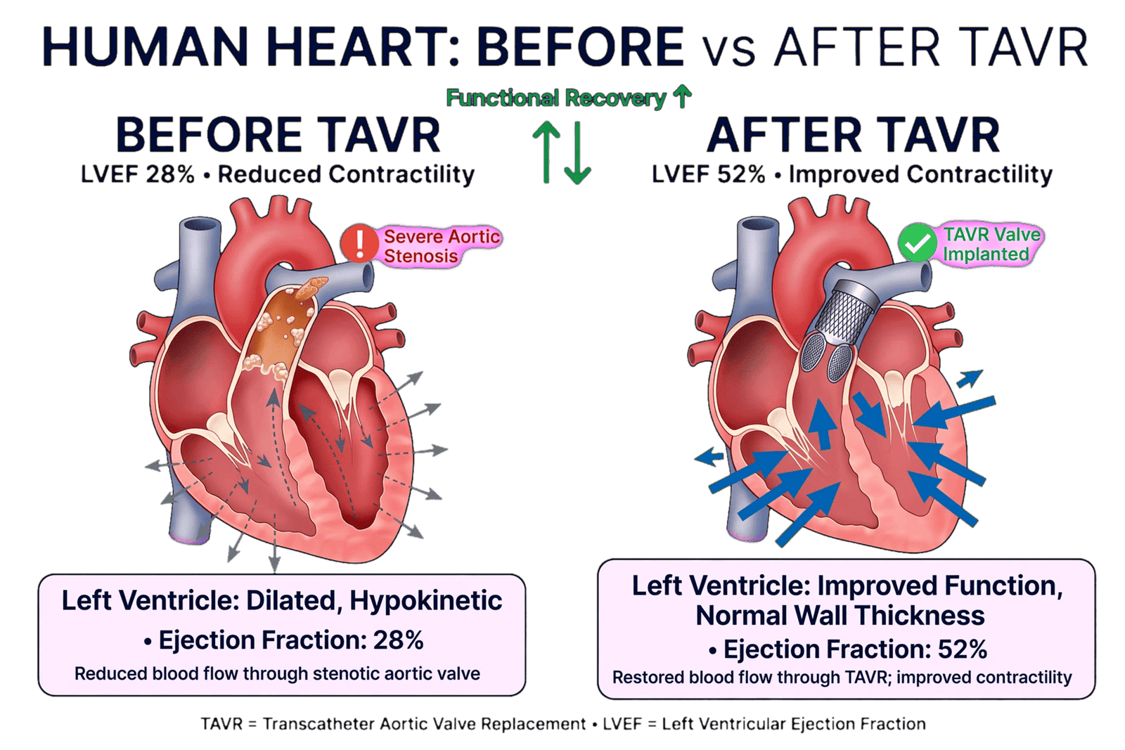
This explains why post-TAVR outcomes are heterogeneous. Some patients demonstrate rapid improvement in LVEF, stroke volume, symptoms, and exercise capacity. Others show only modest ventricular recovery. A third group may have persistent or recurrent heart failure despite apparently normal prosthetic valve function. Contemporary evidence indicates that the degree of left ventricular recovery itself carries prognostic information. In a large multicenter registry of patients with severe LV dysfunction undergoing TAVI, approximately 60% experienced LV recovery, while about 27% achieved normalization of EF above 50%. Importantly, patients without LV recovery had higher three-year mortality.
The concept of “recovery” therefore needs to be separated from “survival” and from “procedural success.” A patient can survive TAVR without meaningful reverse remodeling, and a patient can have excellent prosthetic valve hemodynamics while remaining symptomatic. Conversely, substantial ventricular recovery can occur even in patients with severely reduced baseline EF, demonstrating that a low EF alone should not be interpreted as irreversible myocardial failure.
This distinction has major implications for patient selection and post-procedural surveillance. If recovery depends partly on the reversibility of myocardial disease, then identifying the substrate before TAVR becomes clinically important. The central question changes from “Can the valve be replaced safely?” to “How much of the ventricular disease is still reversible after the valve is replaced?”
The answer is unlikely to come from a single parameter. Baseline EF, transaortic gradient, stroke volume, ventricular dimensions, strain, fibrosis burden, coronary disease, renal function, pulmonary pressures, right ventricular function, atrial rhythm, and concomitant valvular disease all contribute to the biological phenotype.
The contemporary literature therefore supports a more sophisticated concept of TAVR success. Procedural success should be considered the first step; meaningful success requires appropriate hemodynamic correction followed by ventricular and systemic adaptation. Failure of that adaptation may represent persistent myocardial disease rather than failure of the prosthetic valve.
This perspective is particularly relevant in patients with low-flow, low-gradient aortic stenosis and severe LV dysfunction. In such patients, the pressure gradient may underestimate the severity of the valve lesion, while the myocardium may already have advanced fibrosis and impaired contractile reserve. Yet even this group can experience substantial reverse remodeling after intervention. A 2023 study of patients with low-flow, low-gradient AS found that approximately 77% met a predefined criterion for LV reverse remodeling after TAVI, and absence of remodeling was associated with worse clinical outcomes.
Therefore, the central challenge is not simply identifying who can undergo TAVR, but identifying who possesses a sufficiently recoverable myocardium to derive maximal biological benefit. This is the conceptual foundation for modern post-TAVR phenotyping.
The Biology of Left Ventricular Reverse Remodeling After TAVR
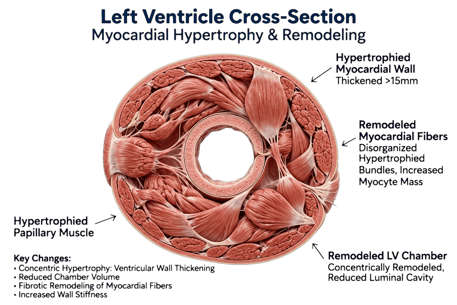
Left ventricular reverse remodeling is one of the most important biological consequences of relieving severe aortic stenosis. Chronic pressure overload forces the ventricle to generate elevated systolic pressures against the obstructed aortic valve. In response, myocardial hypertrophy develops, initially serving as an adaptive mechanism that reduces wall stress. With prolonged exposure, however, hypertrophy becomes associated with interstitial collagen deposition, myocardial fibrosis, impaired relaxation, elevated filling pressures, microvascular dysfunction, and eventually deterioration of systolic mechanics.
TAVR abruptly changes this environment. By reducing transvalvular obstruction, it lowers the pressure gradient between the left ventricle and aorta and decreases the ventricular pressure burden. The myocardium is therefore exposed to a substantially different loading condition immediately after valve replacement. Reverse remodeling can begin rapidly, although its manifestations occur on different time scales.
Some changes occur almost immediately. Hemodynamic unloading may improve forward flow and reduce ventricular pressure. Other changes require weeks or months, including regression of LV mass, improvement in myocardial deformation, reduction in ventricular volumes, and gradual normalization of geometry. The speed and completeness of these changes depend on how much of the pre-existing disease is adaptive and how much has become irreversible.
Cardiac magnetic resonance studies have provided important insight into this process. Early after TAVI, substantial reductions in LV mass can occur, demonstrating that reverse remodeling begins very soon after afterload relief. However, the degree of remodeling differs according to the presence and pattern of myocardial fibrosis. Patients without late gadolinium enhancement have demonstrated greater early LV mass regression than patients with midwall or infarct-pattern fibrosis.
This finding supports a fundamental biological principle: unloading can reverse hypertrophy more readily than it can eliminate established scar. A hypertrophied but relatively viable myocardium may regain more normal geometry after TAVR, whereas a ventricle containing extensive replacement fibrosis has less capacity for functional recovery.
The distinction between interstitial and replacement fibrosis is also important. Diffuse interstitial fibrosis represents expansion of the extracellular matrix and may be partially reversible when the underlying pressure overload is corrected. Replacement fibrosis, particularly dense scar related to ischemia or advanced myocardial injury, is much less likely to recover completely. Contemporary CMR techniques, including late gadolinium enhancement and extracellular volume assessment, may therefore provide information about myocardial reversibility that cannot be obtained from LVEF alone.
Reverse remodeling should also not be defined exclusively by EF. A patient may experience substantial regression of LV mass and improvement in myocardial deformation without a dramatic change in LVEF. Conversely, EF may improve because loading conditions have changed while significant structural abnormalities persist.
This is why longitudinal assessment is essential. Echocardiography after TAVR can track LVEF, LV volumes, LV mass, GLS, diastolic function, left atrial size, and right ventricular performance. CMR can provide more detailed information about myocardial tissue characteristics when clinically appropriate.
The magnitude of reverse remodeling has prognostic significance. Recent reviews have emphasized that reductions in LV mass and improvements in LVEF and GLS after aortic valve intervention are associated with better outcomes, whereas persistent hypertrophy, lack of EF improvement, or impaired strain recovery may indicate residual risk.
However, reverse remodeling is not uniform. Hypertension may maintain afterload even after the valve has been replaced. Diabetes and chronic kidney disease may contribute to myocardial fibrosis and vascular dysfunction. Coronary artery disease may impose an independent ischemic burden. Atrial fibrillation can impair atrial contribution to ventricular filling and promote hemodynamic instability. Pulmonary hypertension and RV dysfunction can limit functional recovery despite satisfactory LV unloading.
This means that TAVR should be conceptualized as removing one major stressor rather than reversing every component of cardiovascular disease.
The term “reverse remodeling” itself can also be misleading if interpreted as complete normalization. Some patients experience partial rather than complete recovery. Persistent LV hypertrophy may remain despite successful valve replacement, and myocardial fibrosis may continue to influence diastolic function and arrhythmia risk.
The clinically important question is therefore not whether reverse remodeling occurs—it usually does to some degree—but whether it is sufficient to restore myocardial function and reduce heart failure burden.
A future approach may classify patients according to remodeling trajectory: rapid responders, gradual responders, partial responders, and nonresponders. Such trajectory-based phenotyping could be more informative than a single postoperative EF measurement.
Ultimately, the biological response to TAVR reflects an interaction between the magnitude of afterload reduction and the reversibility of myocardial injury. The valve determines the hemodynamic correction; the myocardium determines how much of that correction becomes meaningful recovery.
Baseline Predictors of Left Ventricular Recovery: LVEF, Flow, Gradient, and Ventricular Geometry
One of the most clinically relevant questions before TAVR is whether a severely dysfunctional ventricle is likely to recover after valve replacement. Historically, severe LV dysfunction has sometimes been viewed as a marker of irreversible myocardial disease and therefore increased procedural risk. Contemporary evidence challenges this simplistic interpretation.
A low baseline LVEF does not necessarily mean that the ventricle is beyond recovery. In fact, patients with severe LV dysfunction may experience substantial improvement after afterload reduction. In a multicenter registry of 10,872 patients with severe aortic stenosis, 914 had baseline EF ≤30%. Among these patients, 59.5% experienced LV recovery, and 26.7% achieved EF normalization above 50%. The absence of LV recovery, rather than baseline severe dysfunction itself, was associated with worse three-year mortality.
This finding is important because it reverses the usual question. Instead of asking whether a low EF predicts poor outcome, clinicians should ask why the EF is low. If the predominant mechanism is afterload mismatch and potentially reversible myocardial dysfunction, recovery may be substantial. If the mechanism is extensive ischemic scar or advanced fibrosis, recovery is less likely.
Transaortic gradient provides one clue. A higher mean gradient may indicate a more classical high-gradient phenotype in which the ventricle is responding to a true fixed pressure overload. Several studies have found that higher baseline gradients are associated with greater likelihood of LV recovery. In patients with severe LV dysfunction, absence of previous MI, preserved renal function, and mean aortic valve gradient >40 mmHg were associated with greater recovery.
The physiological explanation is plausible: a high-gradient phenotype may represent a ventricle whose dysfunction is more strongly related to valvular afterload, whereas low-gradient disease may be more heterogeneous and may contain a greater proportion of intrinsic myocardial dysfunction.
Low-flow, low-gradient aortic stenosis therefore deserves special consideration. Low forward flow may result from severe LV systolic dysfunction, restrictive physiology, small ventricular cavity, or combined ventricular and vascular abnormalities. In these patients, the gradient alone may underestimate the hemodynamic burden.
Importantly, low flow does not automatically imply poor recovery. A study of 219 patients with low-flow, low-gradient AS undergoing TAVI reported reverse remodeling in approximately 77% and found that lower baseline stroke volume index, very low LVEF, and lower valvulo-arterial impedance were associated with greater likelihood of reverse remodeling.
The relationship between baseline EF and recovery is therefore nonlinear. Patients with very low EF may have a large potential for improvement if afterload mismatch is the dominant mechanism. Conversely, patients with moderately reduced EF may fail to recover if myocardial fibrosis or ischemic injury is advanced.
LV geometry provides additional information. Marked LV dilation may suggest chronic remodeling and a greater degree of structural disease. In a study of patients with reduced EF undergoing transfemoral TAVR, larger LV end-diastolic diameter was independently associated with lower likelihood of early EF improvement.
This supports the concept that duration and severity of myocardial remodeling matter. The ventricle that has remained relatively compact and hypertrophied may be more capable of reverse remodeling than a markedly dilated ventricle with extensive structural deterioration.
Stroke volume is another key parameter. A low stroke volume can indicate advanced ventricular dysfunction, but it can also reflect the interaction between LV contractility, chamber geometry, vascular load, and valvular obstruction. It should therefore be interpreted in conjunction with EF and gradient rather than as an isolated marker.
Valvulo-arterial impedance provides another perspective by estimating the global LV hemodynamic burden created by both valvular and arterial components. This is particularly relevant because replacing the aortic valve does not eliminate systemic arterial hypertension or abnormal vascular stiffness.
Baseline renal function also emerges repeatedly as a predictor of recovery. In the large contemporary registry, estimated GFR >60 mL/min was associated with greater likelihood of LV recovery. Renal dysfunction may represent both a marker and mediator of advanced cardiovascular disease, contributing to volume overload, vascular dysfunction, inflammation, and myocardial fibrosis.
The practical lesson is that pre-TAVR risk assessment should avoid using EF as a binary marker of “recoverable” versus “irreversible” myocardium. Instead, EF should be interpreted as one component of a physiological profile that includes flow, gradient, geometry, renal function, coronary disease, fibrosis, and myocardial mechanics.
This approach allows a more nuanced prediction: a severely impaired ventricle may still be highly recoverable when the dominant pathology is pressure overload, whereas a less severely reduced EF may remain persistently abnormal when structural myocardial injury is advanced.
Myocardial Fibrosis and Global Longitudinal Strain: Identifying the Recoverable and the Irreversible Ventricle
Myocardial fibrosis may be one of the most important biological determinants of ventricular recovery after TAVR. Aortic stenosis produces chronic pressure overload, which stimulates hypertrophic signaling, extracellular matrix expansion, and collagen deposition. Over time, this process can progress from potentially reversible interstitial fibrosis to more advanced replacement fibrosis and scar.
The clinical importance of fibrosis is that valve replacement removes the pressure overload but does not necessarily remove established myocardial scar. Therefore, fibrosis can act as a biological “memory” of the duration and severity of aortic stenosis.
Cardiac magnetic resonance has provided some of the strongest evidence supporting this concept. Late gadolinium enhancement can identify focal replacement fibrosis, while extracellular volume techniques can provide an estimate of diffuse interstitial expansion. In patients undergoing TAVI, greater myocardial fibrosis has been associated with more advanced remodeling and delayed normalization of ventricular geometry and function. In a prospective study using histological myocardial fibrosis, patients with greater fibrosis had worse baseline LV function, more pathological remodeling, delayed recovery, and markedly higher cardiovascular mortality.
Importantly, fibrosis does not necessarily mean that recovery is impossible. The relationship is probabilistic rather than absolute. Patients with substantial fibrosis may still improve after TAVR, but the magnitude and speed of recovery may be reduced.
This distinction becomes clinically relevant when deciding how to interpret a persistent low EF after TAVR. If the patient has extensive fibrosis, persistent dysfunction may represent irreversible myocardial disease. If fibrosis burden is low and valve hemodynamics are excellent, persistent dysfunction may warrant investigation for other reversible causes.
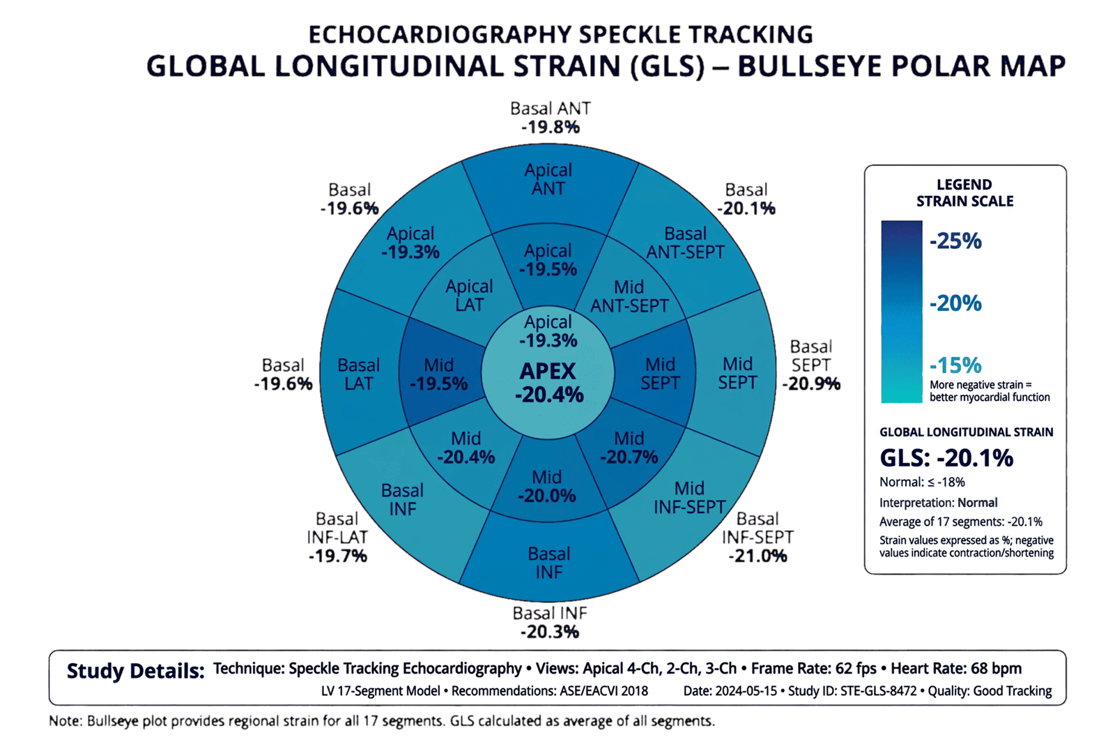
Global longitudinal strain provides a complementary method that is more accessible than CMR in routine practice. GLS measures longitudinal myocardial deformation and can detect systolic dysfunction even when LVEF remains relatively preserved. In the context of TAVR, strain may therefore provide information about myocardial reserve that is not apparent from EF.
Studies have demonstrated that GLS can improve after TAVR, sometimes before major changes in LVEF become evident. One study found that GLS improved at three months while LVEF showed less substantial change, suggesting that myocardial mechanics may recover earlier than conventional chamber-based systolic function.
This creates an important opportunity for early phenotyping. A patient with severe AS and preserved EF but markedly impaired GLS may already have significant myocardial involvement. Such a patient may still benefit from TAVR, but the abnormal strain may indicate a more advanced myocardial phenotype and potentially greater residual risk.
Similarly, postoperative GLS can help distinguish different recovery trajectories. Improvement in GLS may indicate restoration of myocardial mechanics even when EF remains unchanged. Conversely, persistently abnormal or worsening GLS may suggest residual myocardial disease.
Nevertheless, strain should not be treated as a perfect surrogate for fibrosis. GLS is influenced by preload, afterload, blood pressure, heart rate, rhythm, image quality, and software methodology. A low GLS does not prove irreversible fibrosis, just as a normal GLS does not exclude clinically important myocardial pathology.
The most powerful approach may therefore be complementary use of echocardiography and CMR. Echocardiography can provide repeated functional measurements, while CMR can characterize tissue composition when clinically indicated.
CTA-derived extracellular volume is another emerging possibility. A study of patients with impaired LV systolic function undergoing TAVR found that higher extracellular volume was associated with reduced likelihood of early EF recovery. Mean aortic gradient, LV end-diastolic volume, and extracellular volume independently predicted early recovery.
This is particularly interesting because CT is already central to TAVR planning. If tissue characterization can be derived from the same pre-procedural dataset, anatomical planning and myocardial risk stratification could potentially become integrated.
The broader concept is myocardial reserve. Two ventricles with identical EF can have different capacities for recovery because their tissue substrates differ. One may contain viable but hypertrophied myocardium capable of reverse remodeling; the other may contain extensive fibrosis and scar.
The future of TAVR assessment may therefore move toward identifying the “recoverable ventricle” before intervention. This could involve a combination of GLS, CMR fibrosis, CT-derived extracellular volume, LV geometry, flow-gradient phenotype, and clinical history.
Such an approach would not be used to deny TAVR to patients with advanced disease. Rather, it could improve counseling, postoperative surveillance, and interpretation of persistent dysfunction. It could also help identify patients who may need additional heart failure therapy even after successful valve replacement.
The key message is that the valve is mechanical, but the disease is biological. The success of TAVR depends not only on opening the valve but also on whether the myocardium retains sufficient structural and functional reserve to respond to unloading.
Coronary Disease, Prior Myocardial Infarction, and Other Substrates of Persistent LV Dysfunction
Aortic stenosis and coronary artery disease frequently coexist, particularly in older patients undergoing TAVR. This creates a major challenge when interpreting ventricular dysfunction because reduced LVEF may arise from multiple interacting mechanisms rather than isolated valvular afterload.
Prior myocardial infarction is one of the most consistently identified negative predictors of LV recovery after TAVR. In the large multicenter registry of patients with severe LV dysfunction, absence of previous MI was associated with greater likelihood of recovery. Similar findings have been reported in earlier studies of patients with severe LV dysfunction undergoing TAVI.
The explanation is straightforward: infarction produces irreversible loss of cardiomyocytes and replacement fibrosis. Although noninfarcted myocardium can recover after afterload reduction, established scar cannot simply regain contractile function.
The presence of coronary artery disease may therefore create a “dual-hit” model. The ventricle is exposed simultaneously to pressure overload from aortic stenosis and ischemic injury from coronary disease. TAVR corrects the first component but does not automatically correct the second.
This is particularly important in patients with regional wall-motion abnormalities. A globally reduced EF caused by diffuse afterload mismatch may have greater recovery potential than a similarly reduced EF caused by extensive regional ischemic scar.
Coronary assessment before TAVR should therefore be interpreted in the context of ventricular phenotype rather than simply treated as an anatomical exercise. The presence, severity, distribution, and physiological significance of coronary disease may influence both recovery and symptoms.
However, the relationship between coronary revascularization and TAVR outcomes is complex. Not every coronary stenosis is responsible for LV dysfunction, and routine revascularization of every angiographic lesion may not improve outcomes. The important question is whether coronary disease represents an active and clinically meaningful substrate contributing to myocardial dysfunction.
Prior MI is particularly informative because it represents evidence of established myocardial injury rather than simply coronary anatomy.
Other myocardial diseases can similarly limit recovery. Infiltrative cardiomyopathies, inflammatory disease, hypertrophic cardiomyopathy, amyloidosis, and advanced diabetic or hypertensive remodeling may produce ventricular dysfunction that cannot be fully explained by aortic stenosis.
This becomes increasingly important because the population referred for TAVR is heterogeneous. A patient with severe AS and cardiac amyloidosis may have preserved EF but profound diastolic dysfunction and myocardial thickening. Replacing the valve may correct the stenosis but may not eliminate the underlying restrictive physiology.
A similar principle applies to advanced hypertensive heart disease. Even after TAVR, persistent systemic hypertension can maintain LV afterload. If arterial stiffness remains severe, the ventricle may continue to experience a substantial pressure burden despite a normal prosthetic valve.
Diabetes and chronic kidney disease represent additional systemic substrates. Both are associated with endothelial dysfunction, inflammation, myocardial fibrosis, and impaired vascular compliance. Their presence may reduce the probability of complete recovery and may contribute to persistent congestion or exercise intolerance.
Renal dysfunction has repeatedly appeared as a marker of worse recovery. In the contemporary registry, eGFR >60 mL/min was associated with greater likelihood of LV recovery. This does not necessarily imply that renal disease directly prevents myocardial recovery; rather, it may reflect a broader systemic disease burden.
Atrial fibrillation is another important factor. Loss of coordinated atrial contraction can impair LV filling, particularly in a stiff hypertrophied ventricle. AF may therefore contribute to persistent symptoms even when LV systolic function improves.
The interaction between AF and remodeling is bidirectional. Chronic elevation of LV filling pressures promotes atrial enlargement and AF, while AF can worsen hemodynamics and promote further congestion.
Concomitant mitral regurgitation may also influence recovery. Functional MR can improve after TAVR when LV geometry and filling pressures improve, but persistent MR can maintain volume overload and symptoms. In studies of mixed aortic valve disease, postoperative MR and coronary artery disease were independently associated with failure of LV reverse remodeling.
These observations reinforce the concept that post-TAVR heart failure is often multifactorial.
The clinical mistake would be to attribute every persistent symptom to an unsuccessful TAVR. The opposite mistake would be to assume that a normal prosthetic valve excludes cardiac causes of symptoms.
Persistent LV dysfunction after TAVR should therefore trigger a structured search for competing myocardial substrates: prior MI, active ischemia, extensive fibrosis, uncontrolled hypertension, cardiomyopathy, AF, persistent MR, renal disease, and pulmonary or RV disease.
The most useful question is not simply “Did the EF improve?” but “What mechanism is preventing the ventricle from recovering?”
Residual Hemodynamic and Valvular Abnormalities After TAVR: When the New Valve Is Not the End of the Problem
A successful TAVR should substantially reduce the hemodynamic burden imposed by severe aortic stenosis. However, residual obstruction, prosthesis-patient mismatch, paravalvular aortic regurgitation, prosthetic dysfunction, and abnormal blood pressure can limit the expected benefit.
Prosthesis-patient mismatch occurs when the effective orifice area of the implanted prosthesis is too small relative to the patient’s body size. In this situation, the prosthesis may function normally while still generating a relatively high gradient. The problem is therefore not structural valve failure but an inadequate effective valve area for the patient’s flow requirements.
The clinical significance of PPM after TAVR remains an area of active investigation. Contemporary data are more nuanced than earlier surgical literature. A 2026 real-world study of nearly 3,000 patients receiving contemporary self-expanding TAVR valves found predicted PPM in only 5%, with moderate PPM in 3.7% and severe PPM in 1.3%; importantly, PPM was not associated with higher five-year mortality or heart failure hospitalization in that cohort.
This illustrates an important principle: PPM should not automatically be labeled the cause of persistent symptoms. Its clinical significance depends on severity, flow state, patient phenotype, and competing causes of heart failure.
Nevertheless, when a patient has persistent symptoms accompanied by unexpectedly high prosthetic gradients, PPM should be considered. Contemporary guidelines recommend baseline post-procedural echocardiography and ongoing surveillance, with additional imaging when symptoms or signs suggest prosthetic dysfunction.
Paravalvular aortic regurgitation is another potential source of persistent volume loading. Significant regurgitation increases LV volume burden and may counteract the benefits of relieving stenosis. Even when regurgitation is not severe enough to require immediate intervention, it may contribute to incomplete reverse remodeling in susceptible patients.
Residual or progressive mitral regurgitation can be equally important. Functional MR may improve after TAVR as LV pressures and geometry normalize, but it does not always disappear. Persistent MR can maintain elevated left atrial pressure and pulmonary venous hypertension.
The interaction between TAVR and MR is therefore dynamic. Some patients experience rapid improvement, while others remain symptomatic because the mitral valve has become an independent component of the disease.
Right-sided valve disease should also not be ignored. Significant tricuspid regurgitation after TAVR may identify a subgroup with persistent systemic congestion and worse outcomes. In a large TAVR registry, severe or massive TR after TAVR—not simply baseline TR—was associated with increased mortality, and patients considered potential candidates for transcatheter tricuspid intervention had higher risk of persistent severe heart failure symptoms.
Blood pressure represents another residual afterload. TAVR corrects valvular obstruction but does not eliminate systemic arterial hypertension. If arterial pressure remains high, the LV may continue to face increased wall stress. This can blunt reverse remodeling and maintain symptoms.
Valvulo-arterial interaction is particularly important in patients with stiff arteries. The overall burden imposed on the ventricle reflects both the prosthetic valve and systemic vascular load.
Persistent high filling pressures can also occur despite apparently excellent valve hemodynamics. A hypertrophied, stiff ventricle may continue to require high filling pressures even after the valve gradient has normalized.
This explains why a normal mean prosthetic gradient cannot be used as proof that heart failure physiology has resolved.
The appropriate evaluation of persistent symptoms therefore begins with a repeat echocardiographic assessment. The study should include prosthetic valve velocity and gradient, effective orifice area when appropriate, paravalvular regurgitation, LV volumes and EF, diastolic parameters, LA size, RV function, TR severity, and pulmonary pressure estimates.
If the findings remain discordant with the clinical picture, transesophageal echocardiography, cardiac CT, CMR, or invasive hemodynamic assessment may be required depending on the suspected mechanism.
This is particularly important because guidelines emphasize that persistent symptoms after valve intervention warrant evaluation for prosthetic dysfunction, ventricular deterioration, or another cause rather than assuming procedural failure.
Ultimately, the “new valve” should be considered one component of a new cardiovascular system rather than the endpoint of treatment. The post-TAVR ventricle remains sensitive to residual gradients, regurgitation, systemic blood pressure, atrial function, and pulmonary vascular load.
A technically perfect prosthesis cannot compensate for an unresolved hemodynamic phenotype.
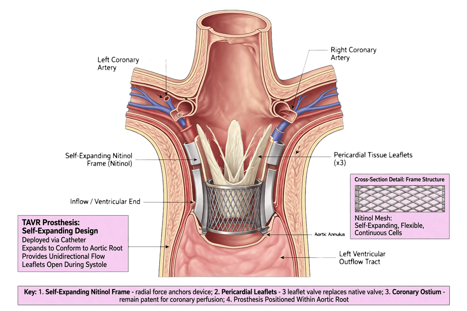
Beyond the Left Ventricle: Atrial Dysfunction, Pulmonary Hypertension, Right Ventricular Failure, and Systemic Comorbidity
Persistent heart failure after TAVR cannot be explained by the LV alone. Severe aortic stenosis produces a chain of hemodynamic consequences extending from the LV to the left atrium, pulmonary circulation, right ventricle, and systemic organs. Removing the valve obstruction interrupts one link in this chain, but downstream abnormalities may persist.
The left atrium is a particularly important marker of chronic disease burden. Long-standing elevation of LV filling pressure causes atrial enlargement and functional deterioration. A large left atrium therefore represents more than a chamber measurement; it can be considered an integrated marker of cumulative exposure to elevated filling pressures.
Even after successful TAVR, left atrial remodeling may persist. If the LV remains stiff, left atrial pressure can remain elevated, especially during exercise. This may explain why some patients have persistent dyspnea despite normalized aortic valve gradients.
Atrial fibrillation further complicates the picture. The loss of atrial contraction may have a disproportionately large impact in patients with hypertrophied and noncompliant ventricles. Persistent AF can therefore maintain symptoms even if LV systolic function improves.
Pulmonary hypertension is another major determinant. Chronic elevation of LV filling pressure can lead to pulmonary venous hypertension and, over time, pulmonary vascular remodeling. In advanced cases, the pulmonary circulation may develop a precapillary component that does not immediately reverse after TAVR.
The right ventricle then becomes exposed to increased afterload. RV dysfunction can perpetuate systemic venous congestion, renal dysfunction, hepatic congestion, peripheral edema, and exercise intolerance.
This creates an important clinical paradox: the LV may improve while the patient remains symptomatic because the RV-pulmonary vascular unit has become an independent disease substrate.
Tricuspid regurgitation often reflects this process. Functional TR may improve after TAVR when pulmonary pressures fall, but severe TR can persist or progress. Post-TAVR severe TR has been associated with worse outcomes, emphasizing that right-sided disease should be reassessed after intervention rather than assumed to resolve automatically.
The concept of ventricular interdependence also matters. RV dilation can shift the interventricular septum and impair LV filling. Thus, advanced RV disease can indirectly affect LV performance.
Systemic comorbidity further complicates recovery. Frailty, chronic kidney disease, diabetes, pulmonary disease, anemia, obesity, and sarcopenia can all contribute to persistent symptoms.
Frailty is especially relevant because improvement in valve hemodynamics does not automatically restore functional capacity. A frail patient may have an anatomically successful procedure but remain limited by skeletal muscle dysfunction, impaired mobility, malnutrition, or multimorbidity. Contemporary real-world data show that frailty remains an important long-term prognostic factor after TAVR.
This is why persistent dyspnea should not automatically be classified as persistent cardiac failure. Pulmonary disease, anemia, deconditioning, obesity, and frailty may mimic or amplify HF symptoms.
Conversely, attributing all symptoms to frailty can also be dangerous because occult residual cardiac disease may be missed.
A structured post-TAVR assessment should therefore distinguish between cardiac and noncardiac limitations. Echocardiography can identify residual cardiac abnormalities, while natriuretic peptides, renal function, hemoglobin, pulmonary evaluation, functional testing, and exercise assessment may be needed when symptoms remain unexplained.
Exercise physiology is particularly valuable in borderline cases. Resting measurements may appear acceptable while exercise reveals abnormal filling-pressure elevation, pulmonary hypertension, or inadequate cardiac output augmentation.
This is especially relevant in HFpEF phenotypes. A patient with preserved EF may have relatively normal resting hemodynamics but develop marked increases in LV filling pressures during exertion. Such patients may remain symptomatic even after technically successful TAVR.
Therefore, post-TAVR recovery should be considered a multichamber and systemic phenomenon.
The clinically meaningful endpoint is not simply normalization of LV EF. It is restoration of adequate forward flow, reduction of filling pressures, improvement in pulmonary vascular load, recovery of RV function, reduction in congestion, and improvement in functional capacity.
This broader perspective also explains why two patients with identical prosthetic valve gradients can have completely different clinical trajectories.
One may have normal atrial and RV function, minimal fibrosis, and good functional reserve. Another may have persistent pulmonary hypertension, severe LA dysfunction, AF, RV impairment, and frailty.
The valve is identical; the cardiovascular phenotype is not.
Persistent Heart Failure After an Apparently Successful TAVR: A Multidimensional Phenotype
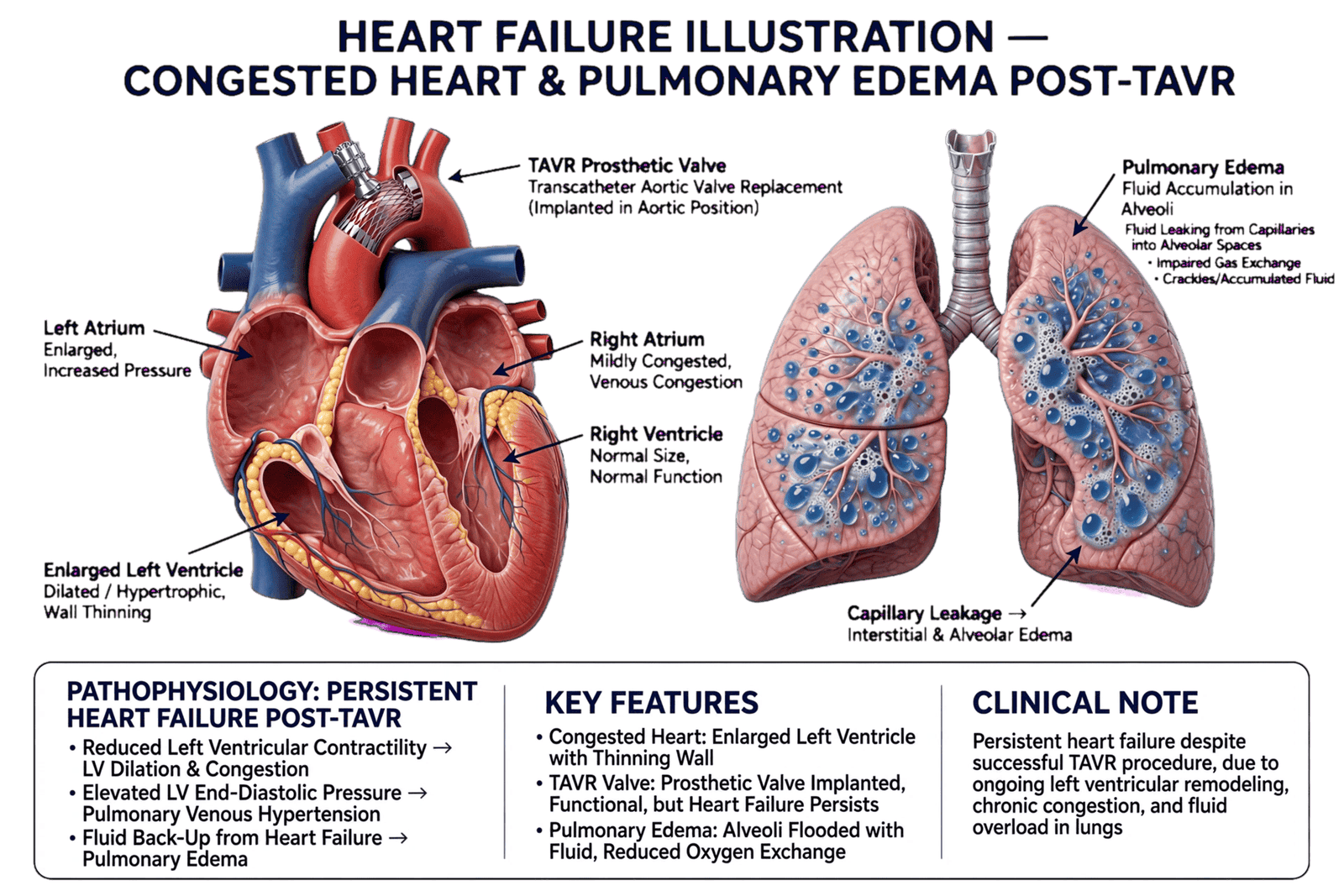
Persistent heart failure after TAVR represents one of the most important unresolved problems in contemporary structural cardiology. The persistence of symptoms despite successful valve implantation challenges the traditional assumption that aortic stenosis is the sole driver of the patient’s cardiovascular syndrome.
The first step in understanding persistent HF is to define what “successful TAVR” actually means. Procedural success should include appropriate valve positioning, acceptable transvalvular gradients, minimal clinically significant regurgitation, and absence of major procedural complications. However, these criteria do not guarantee normalization of ventricular function or filling pressures.
A patient may therefore have procedural success without myocardial recovery.
The mechanisms can be grouped into several overlapping phenotypes.
The first is the irreversible myocardial phenotype. These patients have extensive fibrosis, prior MI, advanced LV remodeling, or another cardiomyopathy. Their valve obstruction has been corrected, but the myocardial substrate has limited recovery potential. CMR fibrosis assessment, GLS, ventricular geometry, and clinical history may help identify this group.
The second is the residual afterload phenotype. Here, the prosthetic valve or systemic circulation continues to impose a meaningful pressure burden. Possible contributors include prosthesis-patient mismatch, residual gradients, systemic hypertension, or vascular stiffness.
The third is the residual volume phenotype. Persistent aortic regurgitation, mitral regurgitation, or tricuspid regurgitation can maintain abnormal loading conditions and prevent complete reverse remodeling.
The fourth is the pulmonary-RV phenotype. These patients may have persistent pulmonary hypertension, RV dysfunction, or severe TR. Their symptoms may be dominated by systemic venous congestion rather than isolated LV dysfunction.
The fifth is the atrial phenotype. Long-standing LA remodeling and AF may sustain elevated filling pressures and exercise intolerance even after LV systolic function improves.
The sixth is the systemic phenotype. CKD, diabetes, obesity, frailty, anemia, pulmonary disease, and deconditioning may contribute substantially to residual symptoms.
Finally, there is a mixed phenotype, which is probably the most common in real-world elderly TAVR populations.
This classification has practical value because it changes the diagnostic strategy.
If the main abnormality is poor LV recovery, the next question should be whether the cause is fibrosis, ischemia, persistent afterload, or inadequate treatment of myocardial disease.
If valve hemodynamics are abnormal, prosthetic evaluation becomes central.
If the valve is normal but pulmonary pressure and TR remain elevated, the focus shifts toward the RV-pulmonary circulation.
If resting echocardiography is relatively reassuring but symptoms persist, exercise hemodynamics or cardiopulmonary exercise testing may reveal abnormalities not visible at rest.
The distinction between persistent HF and persistent symptoms is also essential. Not every patient with dyspnea after TAVR has ongoing congestion. Some have pulmonary disease or severe deconditioning. Conversely, some patients with minimal resting congestion may have significant exercise-induced elevation of filling pressures.
A modern post-TAVR algorithm should therefore integrate clinical symptoms, physical findings, natriuretic peptides, echocardiography, ECG, renal function, and functional capacity.
The echocardiographic component should be multidimensional. It should include prosthetic valve hemodynamics, LV EF, LV volumes, GLS, LV mass, diastolic indices, LA size and possibly strain, RV function, TR severity, and pulmonary pressure estimates.
When these findings are discordant, advanced imaging can clarify the mechanism. CMR may characterize fibrosis and scar. CT can assess prosthetic valve anatomy, leaflet motion, and potentially myocardial extracellular volume in selected protocols. Invasive hemodynamics can distinguish postcapillary from combined pulmonary hypertension and can reveal exercise abnormalities.
The emerging concept is therefore “post-TAVR cardiac phenotyping.”
Rather than reporting simply “EF improved” or “valve functioning normally,” clinicians could describe the patient’s recovery phenotype: complete ventricular recovery, partial reverse remodeling, persistent myocardial dysfunction, residual hemodynamic burden, RV-pulmonary disease, or systemic functional limitation.
This framework is supported by contemporary evidence showing that patients with persistent HF after valve replacement represent a high-risk subgroup. A recent long-term study found that persistent HF before and after aortic valve replacement was associated with markedly worse survival compared with patients without HF, emphasizing that HF after valve replacement is not a benign residual symptom complex.
The clinical implications are substantial. Patients predicted to have incomplete recovery may require closer follow-up, more aggressive management of hypertension and coronary disease, optimization of guideline-directed HF therapy where indicated, evaluation for residual valve disease, and consideration of advanced testing.
Importantly, the existence of a normal prosthetic valve should not end the diagnostic process.
The ultimate objective of TAVR is not merely to implant a valve. It is to restore a cardiovascular system capable of delivering adequate flow at acceptable filling pressures and supporting functional independence.
Therefore, the future definition of TAVR success should evolve from a procedural endpoint to a biological endpoint.
The key question after TAVR should be: “Has the patient’s cardiovascular phenotype actually recovered?”
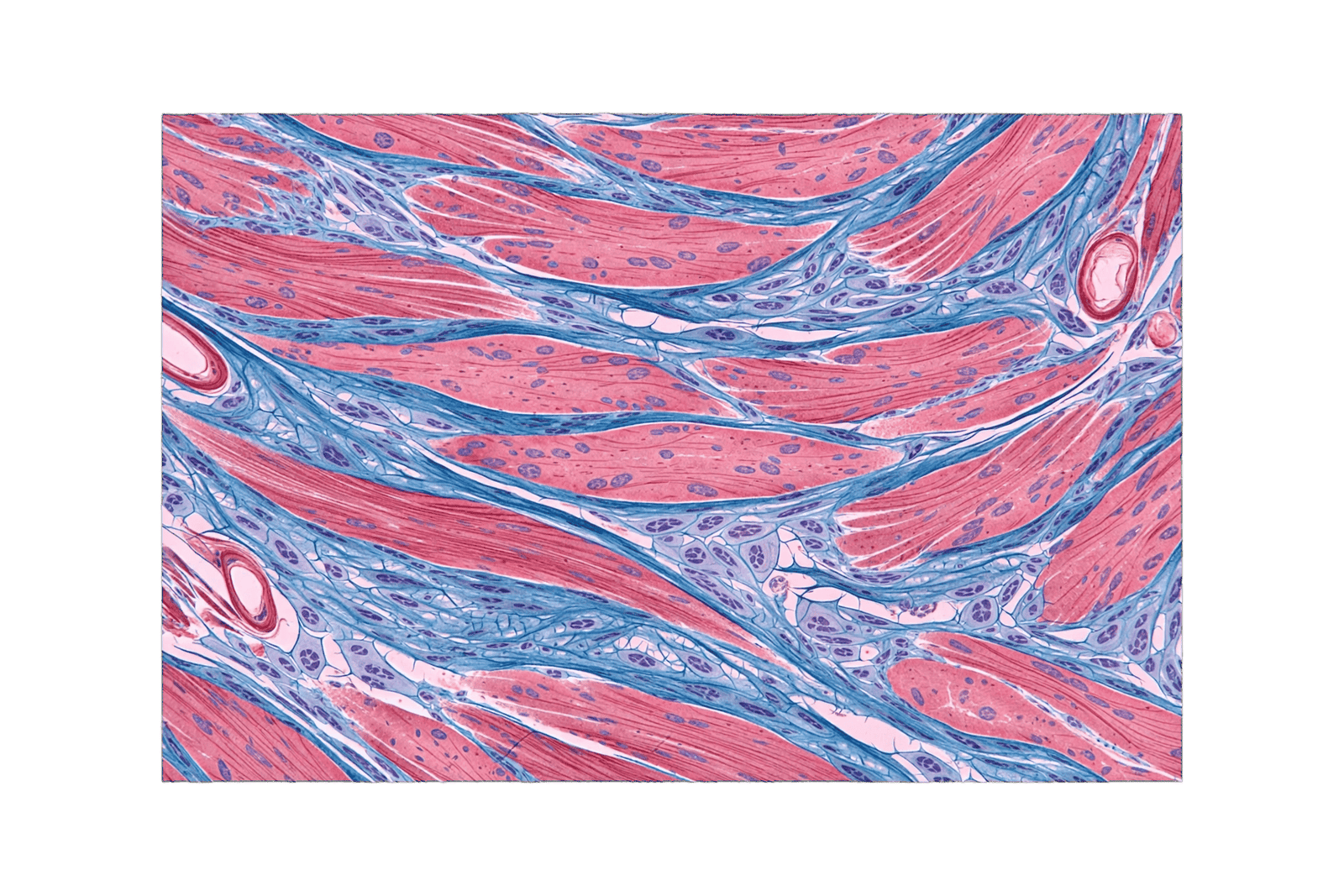
Clinical Risk Stratification and the Future of Precision TAVR Follow-Up
The growing recognition of heterogeneous ventricular recovery after TAVR creates an opportunity to redesign post-procedural risk stratification. Traditional follow-up focuses heavily on valve function, symptoms, and LVEF. These remain indispensable, but they may not adequately identify patients at risk of persistent heart failure.
A precision follow-up strategy would begin before TAVR by establishing a detailed baseline phenotype.
The baseline assessment should include LVEF, LV volumes, LV mass, GLS where feasible, stroke volume index, mean transaortic gradient, valve area, valvulo-arterial impedance, LA size, RV function, TR, pulmonary pressure, renal function, coronary disease, prior MI, rhythm, and frailty.
Advanced imaging could then be selectively added. CMR may be particularly valuable when the distinction between recoverable and irreversible myocardial disease is clinically important. CT-derived extracellular volume is an emerging approach that may eventually provide additional information without requiring a separate CMR examination.
The second component is early post-TAVR reassessment.
A technically successful procedure should be followed by a baseline post-procedural echocardiogram. Current guidelines recommend an initial post-intervention TTE and ongoing surveillance, with repeat imaging prompted by changes in symptoms or examination; annual imaging is considered reasonable for transcatheter bioprosthetic valves.
But future surveillance may become more sophisticated than simply repeating EF and valve gradients.
The trajectory of GLS, LV mass regression, LV volumes, LA mechanics, and RV function could provide a multidimensional recovery profile.
For example, a patient may demonstrate:
- normal prosthetic valve function;
- rapid improvement in GLS;
- reduction in LV mass;
- stable or improving EF;
- decreasing LA pressure surrogates.
This would represent a favorable remodeling trajectory.
Another patient might demonstrate:
- normal prosthetic gradients;
- persistent severely impaired GLS;
- minimal LV mass regression;
- persistent LA enlargement;
- pulmonary hypertension.
This phenotype should trigger investigation for advanced myocardial or pulmonary vascular disease even if the prosthesis is functioning normally.
AI and machine learning may eventually make such multidimensional phenotyping scalable. Algorithms can integrate dozens of echocardiographic variables and potentially analyze raw echocardiographic videos rather than relying exclusively on manually measured parameters.
However, AI should not be considered a substitute for clinical reasoning. Its role should be to identify patterns, quantify measurements, improve reproducibility, and estimate risk while providing interpretable information.
The future may involve an AI-generated “post-TAVR recovery score” incorporating baseline myocardial substrate, procedural hemodynamics, early LV response, and clinical comorbidity. Such a score could identify patients requiring intensified follow-up.
Yet this remains a research objective rather than an established standard. Predictive accuracy alone is insufficient. A model must demonstrate external validity and, ideally, show that acting on its predictions improves patient outcomes.
Another major research priority is determining whether early changes in myocardial mechanics predict long-term outcomes better than conventional EF. If GLS improves before EF, then early strain recovery could potentially become an intermediate endpoint for evaluating ventricular response.
Similarly, CMR fibrosis and CT-derived extracellular volume may help identify patients unlikely to achieve full recovery before TAVR. This could improve patient counseling and postoperative expectations.
Therapeutic implications are equally important. Persistent HF after TAVR should not be treated as a single disease. Management should be phenotype-specific.
Patients with HFrEF and persistent systolic dysfunction may require optimization of guideline-directed medical therapy where appropriate.
Patients with uncontrolled hypertension require aggressive blood-pressure management.
Patients with residual MR or TR may require valve-team reassessment.
Patients with persistent pulmonary hypertension may require characterization of the underlying hemodynamic mechanism.
Patients with AF require appropriate rhythm and rate management and stroke prevention according to established indications.
Patients with frailty and deconditioning may benefit from rehabilitation and nutritional intervention.
This represents a transition from “post-TAVR follow-up” to “post-TAVR cardiovascular rehabilitation.”
The concept of recovery should also include functional endpoints. Improvement in NYHA class, walking distance, quality of life, exercise capacity, and independence may be more meaningful to patients than a modest numerical change in EF.
A future precision TAVR pathway could therefore include three stages:
Stage 1: pre-procedural myocardial characterization.
Stage 2: early assessment of hemodynamic and mechanical response.
Stage 3: longitudinal assessment of structural, functional, and clinical recovery.
This approach recognizes that TAVR changes the valve immediately but changes the patient gradually.
The most important unanswered question is whether earlier identification of nonresponders can change outcomes. If persistent myocardial dysfunction is recognized early, clinicians may be able to intervene before progressive HF becomes established.
Thus, the next frontier is not simply better valve technology. It is better understanding of the patient who receives the valve.
Conclusions: From Procedural Success to Biological Recovery
TAVR has become one of the most effective interventions for severe aortic stenosis, but the success of valve replacement should no longer be judged exclusively by procedural endpoints. The ultimate clinical objective is restoration of cardiac function and reduction of heart failure burden.
Left ventricular reverse remodeling is a major mechanism through which TAVR produces clinical benefit. Relief of pressure overload can lead to regression of hypertrophy, improvement in myocardial mechanics, reduction in ventricular volumes, and recovery of LVEF. However, the extent of this response varies substantially between patients.
The most important determinant is likely the biological state of the myocardium at the time of intervention.
Patients with predominantly afterload-mediated dysfunction may experience dramatic recovery. Patients with extensive fibrosis, prior myocardial infarction, advanced ventricular remodeling, or competing cardiomyopathies may have incomplete recovery despite excellent prosthetic valve function.
Baseline LVEF should therefore not be interpreted in isolation. Severe LV dysfunction can recover substantially, and contemporary registry data demonstrate that more than half of patients with EF ≤30% may experience meaningful recovery after TAVR. Conversely, the absence of recovery identifies a high-risk phenotype.
Flow and gradient are also important. High-gradient disease and the absence of previous MI appear to favor recovery, while low-flow states require nuanced interpretation rather than automatic classification as irreversible disease.
Myocardial fibrosis provides a deeper biological explanation. CMR and emerging CT tissue-characterization techniques suggest that greater fibrosis burden is associated with delayed or impaired ventricular recovery. GLS offers a practical echocardiographic window into myocardial mechanics and may detect dysfunction or recovery before conventional EF changes become obvious.
Persistent heart failure after TAVR must also be approached as a multichamber syndrome. Residual MR, TR, pulmonary hypertension, RV dysfunction, AF, persistent LA dysfunction, renal disease, hypertension, diabetes, frailty, and pulmonary disease can all contribute to continued symptoms.
The prosthetic valve can therefore be completely successful while the cardiovascular syndrome remains incompletely corrected.
This leads to a more meaningful definition of TAVR success:
Procedural success is valve replacement. Biological success is recovery of the cardiovascular system.
The future of TAVR follow-up should consequently move toward multidimensional phenotyping. LVEF, GLS, LV geometry, myocardial fibrosis, flow-gradient physiology, prosthetic valve hemodynamics, atrial function, RV-pulmonary interaction, and systemic comorbidity should be interpreted together.
The objective should not be to generate more measurements for their own sake. Rather, the objective is to determine which abnormalities remain, whether they are reversible, and what clinical action should follow.
A patient with persistent HF after TAVR should therefore not automatically be classified as a procedural failure. The clinician should ask whether the dominant problem is irreversible myocardial disease, residual hemodynamic burden, pulmonary-RV dysfunction, atrial disease, ischemia, or systemic limitation.
This approach also changes patient counseling. Before TAVR, patients can be informed that valve replacement will correct the obstruction but that ventricular recovery depends on the underlying myocardial substrate. After TAVR, persistent dysfunction should be evaluated systematically rather than attributed to age or frailty without evidence.
Future research should focus on prospective validation of recovery predictors, standardized use of strain, tissue characterization, AI-assisted phenotyping, and clinical trials testing whether intervention based on early recovery trajectories improves outcomes.
The most important conceptual shift is therefore simple but profound: the end point of TAVR should not be the moment the prosthesis is implanted. It should be the degree to which the heart, lungs, circulation, and patient recover afterward.
Beyond procedural success, the real question is whether the myocardium can recover—and, when it cannot, whether we can identify the reason early enough to change the patient’s trajectory.