



Introduction: Why Ejection Fraction Alone Is No Longer Enough
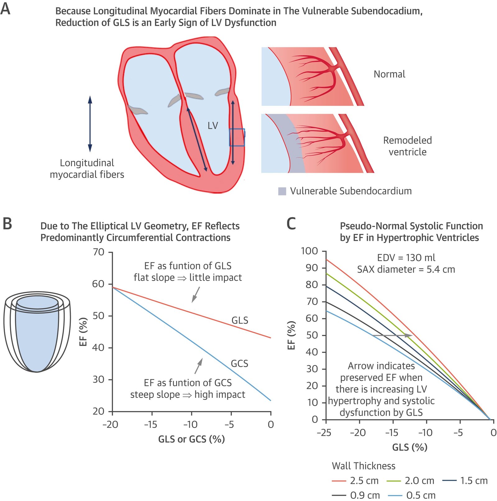
Heart failure has traditionally been characterized and clinically classified through the measurement of left ventricular ejection fraction (LVEF). LVEF remains fundamental because it provides clinically useful information, contributes to heart failure classification, and influences therapeutic decision-making. However, an increasing body of evidence demonstrates that LVEF is an incomplete surrogate for myocardial function. Ejection fraction describes the proportion of ventricular end-diastolic volume expelled during systole, but it does not directly quantify myocardial fiber deformation, regional contractile behavior, ventricular–arterial interaction, filling pressures, myocardial stiffness, or the complex interactions between the left ventricle, right ventricle, left atrium, pulmonary circulation, and systemic circulation.
The limitations of LVEF become particularly important in patients with early myocardial disease and in those with heart failure with preserved or mildly reduced ejection fraction. A patient may have an apparently normal LVEF while already demonstrating impaired myocardial deformation, abnormal diastolic reserve, increased filling pressures, left atrial remodeling, or right ventricular dysfunction. Conversely, two patients with the same LVEF may have substantially different myocardial phenotypes, etiologies, prognoses, and therapeutic responses. Consequently, contemporary heart failure imaging is progressively moving from a single-number model toward multidimensional phenotyping.
The American Heart Association/American College of Cardiology/Heart Failure Society of America guideline recognizes the importance of echocardiography for structural and functional assessment and specifically notes that myocardial deformation indices such as global longitudinal strain may identify subclinical LV systolic dysfunction. Contemporary European imaging literature similarly emphasizes that strain provides information complementary to LVEF and may reveal abnormalities that remain concealed when ventricular function is assessed solely through conventional volumetric parameters.
This conceptual shift is important because heart failure is not an abrupt transition from normal cardiac function to overt ventricular failure. It is often a continuum involving molecular injury, altered myocardial mechanics, impaired relaxation, increasing stiffness, neurohormonal activation, ventricular and atrial remodeling, pulmonary vascular abnormalities, and eventually clinical congestion and exercise intolerance. Imaging capable of identifying earlier stages of this continuum may therefore have greater clinical value than imaging that becomes abnormal only after substantial structural deterioration.
Myocardial strain represents one of the most important developments in this context. Rather than asking only how much blood the ventricle ejects, strain asks how the myocardium deforms during the cardiac cycle. Three-dimensional echocardiography adds a more complete representation of ventricular geometry and volume, while artificial intelligence introduces the possibility of automated measurements, pattern recognition, and integration of complex imaging information.
Importantly, these technologies should not be interpreted as competing alternatives. Their greatest potential lies in integration. Strain can characterize myocardial mechanics; three-dimensional imaging can characterize chamber geometry and volumes; Doppler and conventional echocardiography can estimate filling pressures and hemodynamics; and artificial intelligence can potentially combine these variables with clinical and multimodal imaging data.
The emerging paradigm is therefore not “strain instead of EF” or “AI instead of echocardiographers.” It is a transition toward a richer cardiac phenotype in which LVEF becomes one component of a broader quantitative assessment. Such an approach may improve early detection, refine prognosis, distinguish disease mechanisms, and ultimately support more individualized treatment.
Understanding Myocardial Strain: From Chamber Emptying to Myocardial Deformation
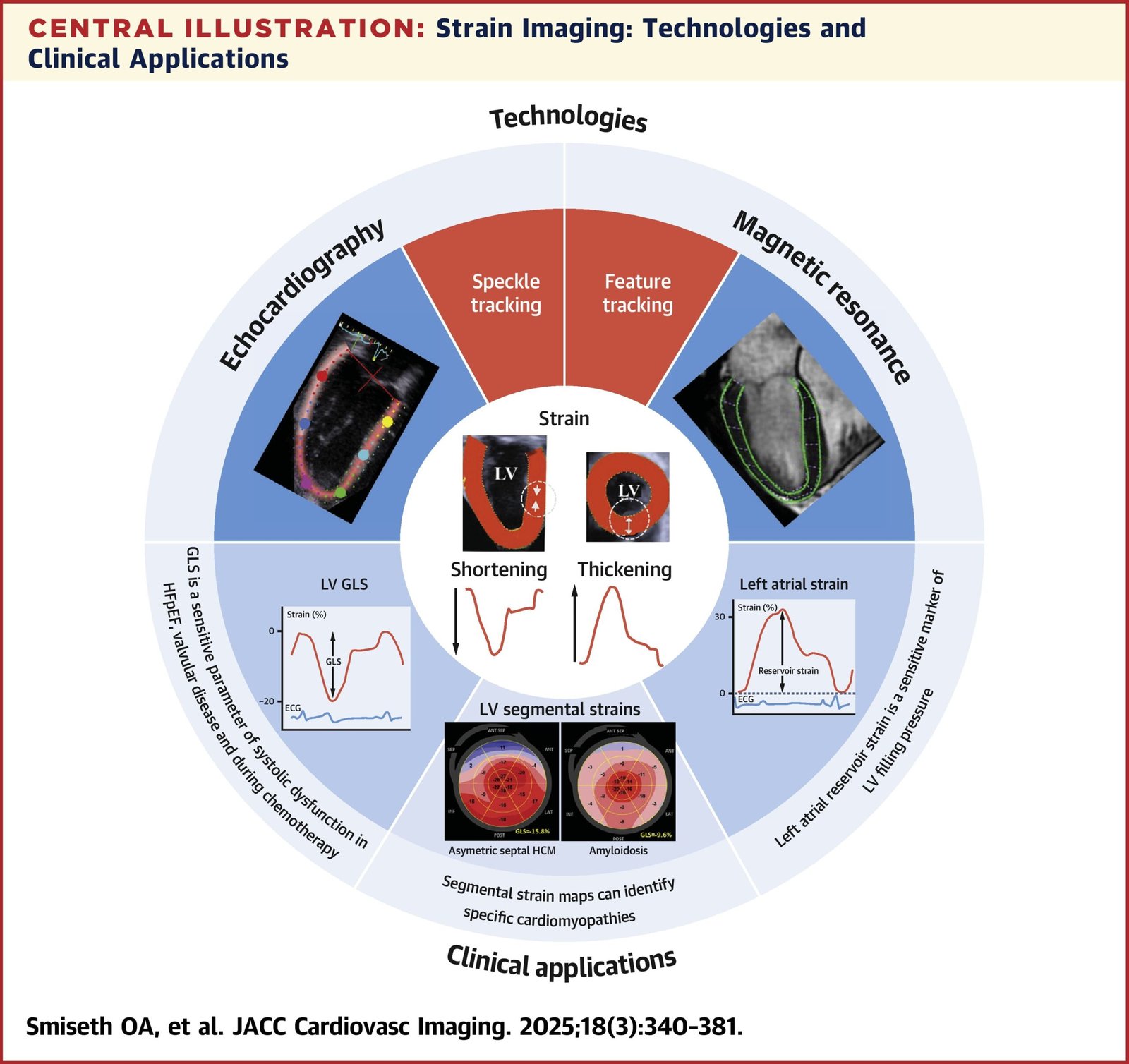
Myocardial strain is a measure of tissue deformation relative to its original dimension. In the heart, deformation occurs in multiple directions, reflecting the complex architecture of myocardial fibers. Longitudinal shortening, circumferential shortening, radial thickening, twisting, and untwisting all contribute to effective ventricular contraction and relaxation. Conventional LVEF captures the integrated result of these processes at the chamber level, whereas strain attempts to characterize the mechanics producing that result.
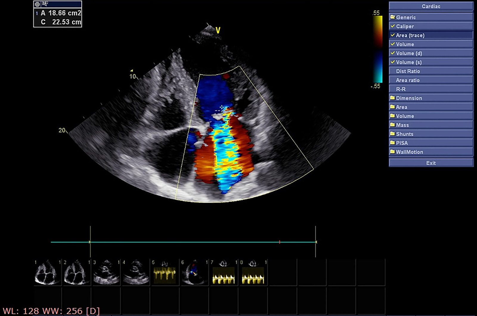
Speckle-tracking echocardiography has become a major clinical method for assessing myocardial deformation. It tracks naturally occurring acoustic markers, or speckles, within the myocardium throughout the cardiac cycle. This permits estimation of deformation without relying exclusively on Doppler-derived velocity measurements. Among the available parameters, global longitudinal strain (GLS) has attracted the greatest clinical attention because longitudinal myocardial fibers can become dysfunctional relatively early in several cardiac diseases.
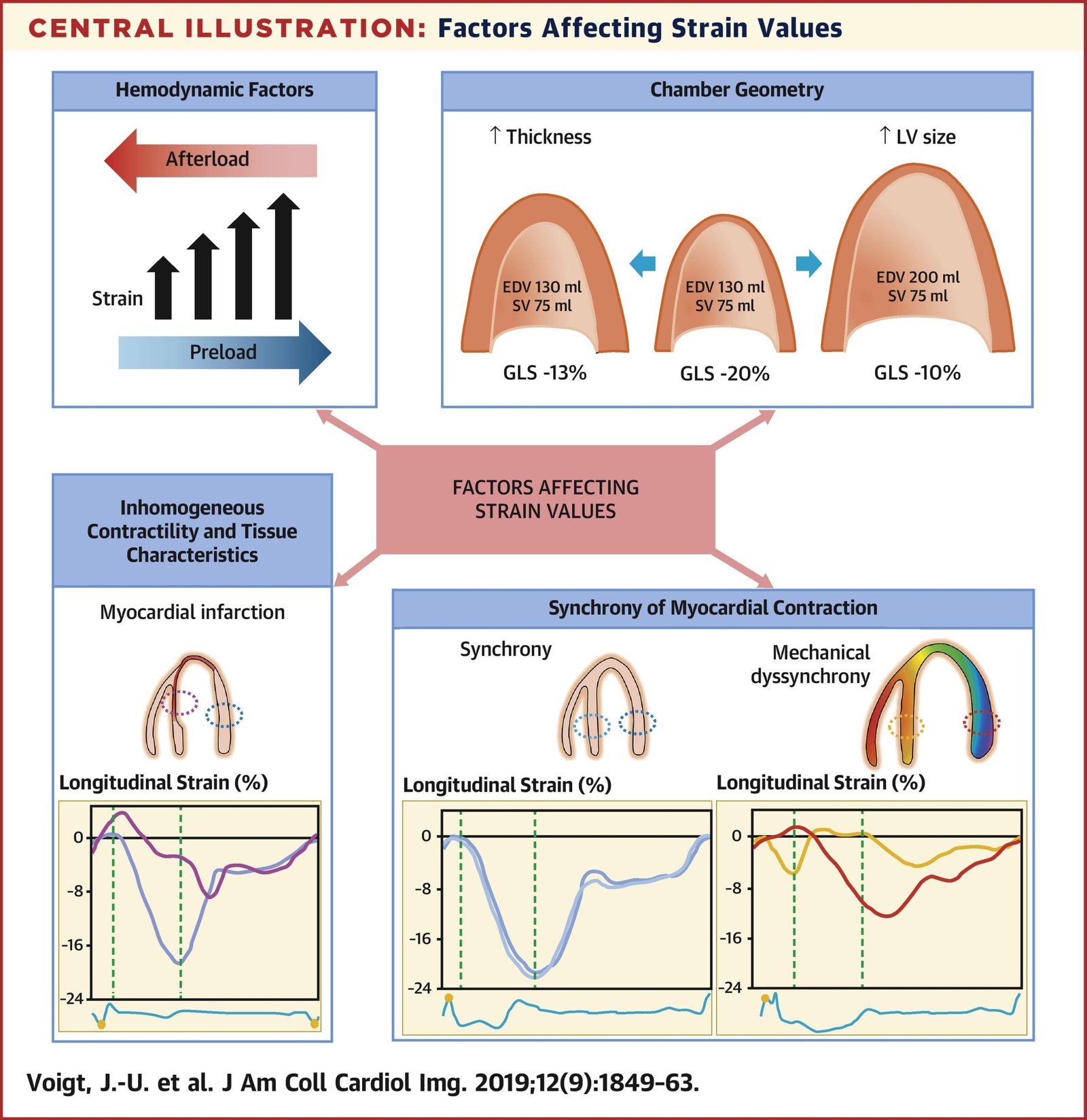
The interpretation of GLS requires an important conceptual distinction. More negative values generally indicate greater longitudinal shortening, whereas less negative values indicate impaired deformation. Therefore, a reduction in the absolute magnitude of GLS represents worsening longitudinal systolic function. However, a universal threshold should not be applied indiscriminately because strain values depend on vendor-specific software, image quality, loading conditions, age, sex, heart rate, rhythm, acquisition technique, and analysis methodology.
The biological rationale for the sensitivity of GLS is closely related to myocardial architecture. Subendocardial longitudinal fibers may be particularly vulnerable to ischemia, fibrosis, increased wall stress, inflammation, and other pathological processes. Longitudinal dysfunction can therefore precede a measurable decline in global pump performance. This explains why patients with apparently preserved LVEF may nevertheless demonstrate abnormal GLS.
Strain also offers spatial information. Segmental strain maps can reveal heterogeneous myocardial dysfunction that may suggest specific disease mechanisms. Certain cardiomyopathies produce characteristic patterns of regional deformation, while ischemic disease may produce abnormalities corresponding to coronary territories. In systemic diseases, strain can provide a quantitative window into myocardial involvement before conventional parameters become clearly abnormal.
Another important advantage is that strain can be evaluated across different chambers. Left ventricular GLS remains the most established parameter, but right ventricular longitudinal strain and left atrial strain are increasingly studied. These measurements expand cardiac assessment beyond isolated LV systolic performance and may better capture the systemic and multichamber nature of heart failure.
Nevertheless, strain is not a direct measurement of contractility in isolation. Myocardial deformation is influenced by preload, afterload, ventricular geometry, blood pressure, and loading conditions. A reduced GLS therefore should not automatically be interpreted as irreversible myocardial damage. Instead, it should be interpreted within the clinical and hemodynamic context.
Reproducibility is another important consideration. Differences between vendors, software platforms, acquisition protocols, frame rates, image quality, and analysis algorithms can influence measured strain values. Contemporary implementation therefore requires standardized acquisition and reporting procedures and awareness of inter-vendor variability.
The significance of strain is ultimately not that it creates another number for the echocardiography report. Its importance lies in its ability to move cardiac imaging from a purely chamber-based description toward a more mechanistic representation of myocardial behavior. This makes strain particularly attractive for early disease detection, risk stratification, phenotyping, and longitudinal assessment.
Global Longitudinal Strain in Early Detection and Risk Stratification of Heart Failure
The clinical appeal of GLS is strongest when conventional LVEF appears normal or only mildly abnormal. In these patients, the identification of subtle systolic dysfunction may provide information that is otherwise unavailable from standard echocardiography. Multiple studies have demonstrated that abnormal GLS is associated with adverse outcomes across different cardiovascular conditions, and meta-analytic evidence suggests that GLS can provide prognostic information beyond LVEF.
In patients with HFpEF, this distinction is especially important. HFpEF is not synonymous with normal myocardium. Preserved EF can coexist with impaired longitudinal shortening, increased myocardial stiffness, left ventricular hypertrophy, atrial dysfunction, abnormal ventricular–arterial coupling, pulmonary hypertension, and impaired chronotropic or contractile reserve. GLS can therefore expose an element of systolic myocardial dysfunction hidden behind preserved global chamber ejection.
Meta-analytic evidence has demonstrated significantly lower GLS values in patients with HFpEF compared with healthy and asymptomatic populations. Importantly, abnormal GLS has also been associated with adverse cardiovascular outcomes in HFpEF. This supports the concept that preserved EF should not be interpreted as evidence that systolic myocardial mechanics are entirely normal.
The prognostic value of GLS extends beyond HFpEF. In acute and chronic heart failure populations, GLS has been associated with mortality and hospitalization independently of LVEF. This is clinically meaningful because LVEF can remain relatively stable while myocardial mechanics deteriorate. Serial GLS may therefore provide a complementary method for monitoring disease progression or response to treatment.
However, the clinical interpretation of GLS should remain cautious. A single abnormal value should not automatically trigger a diagnosis of heart failure in an asymptomatic patient. Heart failure is a clinical syndrome, and imaging findings must be interpreted alongside symptoms, physical findings, natriuretic peptides, structural abnormalities, filling pressure estimates, rhythm, comorbidities, and other diagnostic data.
The concept of “early detection” should therefore be defined carefully. Strain can identify subclinical myocardial dysfunction, but subclinical dysfunction is not equivalent to symptomatic heart failure. The clinical objective is to identify individuals at elevated risk or with early cardiac involvement who may benefit from closer evaluation and preventive intervention—not to label every abnormal strain measurement as overt HF.
GLS may also be useful in populations exposed to myocardial injury from systemic disease or therapy. In cardio-oncology, for example, strain has been investigated for detecting early myocardial dysfunction before an overt reduction in LVEF becomes apparent. Similar principles may apply to hypertensive heart disease, diabetes, valvular disease, infiltrative disease, ischemic heart disease, and other conditions associated with progressive myocardial remodeling.
An additional advantage is that GLS can potentially function as a longitudinal biomarker. Changes over time may be more informative than isolated values. A patient whose LVEF remains within a conventional normal range but demonstrates progressive deterioration in GLS may represent a different biological trajectory from a patient with stable mechanics.
Yet serial strain assessment also introduces challenges. Measurement variability can mimic biological change. Therefore, meaningful longitudinal interpretation requires consistency in imaging platform, acquisition quality, analysis software, loading conditions, and clinical context. Small numerical changes should not be overinterpreted without considering measurement error and reproducibility.
The strongest future application of GLS may therefore be as one component of a multiparametric risk model. Combining GLS with LVEF, LV volumes, LV mass, left atrial volume, E/e′, tricuspid regurgitation velocity, right ventricular function, natriuretic peptides, clinical risk factors, and potentially AI-derived imaging features may provide substantially richer phenotyping than any single measurement.
Strain-Based Phenotyping Across HFrEF, HFmrEF, and HFpEF
The traditional classification of heart failure according to LVEF remains clinically useful, but it does not fully capture the biological heterogeneity within each EF category. HFrEF, HFmrEF, and HFpEF contain multiple phenotypes generated by different combinations of ischemic injury, pressure overload, volume overload, fibrosis, inflammation, metabolic disease, infiltrative processes, genetic cardiomyopathy, valvular disease, atrial dysfunction, and pulmonary vascular disease.
Strain imaging provides an opportunity to examine these groups from a mechanistic perspective. In HFrEF, markedly impaired GLS generally accompanies reduced global systolic performance, but the relationship between LVEF and myocardial mechanics is not perfectly linear. Two patients with similar EF may have different distributions of regional dysfunction, different ventricular geometry, and different degrees of residual viable myocardium.
In HFmrEF, strain may be particularly informative because this group occupies an intermediate region where the boundaries between phenotypes are biologically blurred. A moderately reduced EF can result from previous myocardial infarction, dilated cardiomyopathy, tachycardia-mediated dysfunction, valvular disease, hypertension, or transient myocardial injury. Strain patterns may provide additional clues regarding the underlying mechanism.
HFpEF presents an even greater challenge. The preserved EF phenotype includes patients with hypertensive remodeling, obesity-related cardiometabolic disease, atrial fibrillation, pulmonary hypertension, renal dysfunction, infiltrative disease, valvular disease, and other systemic conditions. These patients may have different degrees of LV longitudinal dysfunction, left atrial impairment, RV involvement, and pulmonary vascular abnormalities.
Segmental strain patterns may also support etiological phenotyping. Although no strain pattern should be treated as a standalone diagnostic test, regional deformation can contribute to recognition of specific cardiomyopathies. For example, disproportionate regional impairment may prompt consideration of ischemic disease or specific myocardial disorders and may help guide subsequent CMR, genetic evaluation, or other investigations.
The right ventricle deserves special consideration. Conventional measures such as TAPSE and fractional area change remain clinically important, but RV longitudinal strain can provide additional information regarding myocardial deformation. In HFpEF, impaired RV mechanics may identify patients with more advanced pulmonary vascular disease or worse prognosis. This supports a transition from an LV-centered model toward a biventricular and multichamber model.
Left atrial strain provides another potentially important dimension. The left atrium functions as a reservoir, conduit, and booster pump, and its mechanics are influenced by LV filling pressures and atrial remodeling. Abnormal left atrial strain may therefore provide information regarding chronic pressure exposure and atrial dysfunction that is not captured fully by atrial volume alone.
The concept of phenotyping should consequently move beyond labels such as HFrEF or HFpEF. A more informative profile might describe a patient according to LV systolic mechanics, ventricular geometry, diastolic function, atrial function, RV performance, pulmonary pressures, valvular abnormalities, and systemic context.
This approach has potential therapeutic implications. Heart failure therapies are increasingly individualized according to phenotype and comorbidity rather than EF alone. Imaging biomarkers may help identify patients with particular biological characteristics, although most strain-based therapeutic decisions remain investigational and should not be presented as established treatment-selection rules without supporting outcome evidence.
An important research opportunity is to determine whether strain-defined phenotypes predict differential responses to therapy. If specific patterns of mechanical dysfunction consistently identify patients who benefit from particular interventions, strain could move from a prognostic biomarker toward a treatment-guiding biomarker.
At present, however, this remains an evolving field. The correct position is that strain enhances phenotyping and risk assessment, but it does not yet replace comprehensive clinical evaluation or guideline-based classification. The future likely lies in combining strain with clinical, biochemical, structural, hemodynamic, and molecular information.
Three-Dimensional Echocardiography: Advancing Volumetric and Structural Phenotyping
Two-dimensional echocardiography remains the foundation of routine cardiac imaging, but its geometric assumptions can introduce limitations when ventricular shape becomes abnormal. Three-dimensional echocardiography offers a more direct representation of cardiac chambers and can improve measurement of ventricular volumes and ejection fraction by reducing reliance on geometric assumptions.
The principal value of three-dimensional echocardiography in heart failure is therefore not simply that it produces visually impressive images. Its deeper value lies in quantitative volumetric characterization. Accurate LV end-diastolic and end-systolic volumes are essential for understanding ventricular remodeling, and changes in these parameters can provide information about disease progression and reverse remodeling.
Three-dimensional imaging can be particularly useful when ventricular geometry is complex. Dilated ventricles, regional wall-motion abnormalities, aneurysmal remodeling, and abnormal chamber shape may challenge conventional two-dimensional methods. By capturing the chamber as a volumetric dataset, 3D echocardiography can provide a more comprehensive assessment of ventricular architecture.
This has implications for heart failure phenotyping. A patient with a moderately reduced EF and severe LV dilation represents a different phenotype from a patient with the same EF and relatively preserved ventricular dimensions. Similarly, changes in LV volumes following medical or device therapy may reveal reverse remodeling even when changes in EF appear modest.
Three-dimensional echocardiography can also improve assessment of the right ventricle, a chamber that is difficult to characterize accurately with conventional two-dimensional geometry because of its complex crescent-shaped anatomy. Three-dimensional RV volumes and ejection fraction can provide more comprehensive structural information, although image quality and temporal resolution remain important limitations.
Another potential application is integration with strain. Three-dimensional speckle-tracking approaches attempt to quantify deformation in multiple directions from a volumetric dataset. This could provide information about longitudinal, circumferential, radial, and area deformation within a unified three-dimensional framework.
However, 3D strain remains less standardized than conventional 2D GLS. Acquisition can be affected by lower temporal resolution, stitching artifacts, irregular rhythm, respiratory motion, and limited acoustic windows. Therefore, promising technical capability should not automatically be equated with established clinical superiority.
Three-dimensional echocardiography also has value in structural heart disease, where accurate assessment of valves and chambers may influence the diagnosis and procedural strategy. In patients with heart failure associated with valvular disease, precise quantification of ventricular remodeling and valve anatomy may be particularly important.
The relationship between 3D imaging and LVEF is therefore complementary rather than adversarial. The goal is not to eliminate EF but to measure it more accurately while simultaneously capturing the structural context in which EF exists.
A major future opportunity is the creation of integrated 3D datasets containing anatomy, volumes, motion, and deformation. These datasets are particularly suitable for computational analysis because they contain substantially more information than isolated measurements. Artificial intelligence may ultimately use these volumetric datasets to identify subtle phenotypes that are difficult to describe manually.
Nevertheless, implementation barriers remain. Three-dimensional acquisition requires appropriate equipment, operator expertise, adequate image quality, and efficient analysis workflows. In busy clinical environments, acquisition and post-processing time may limit adoption. Automated segmentation and AI-assisted analysis may reduce this burden, potentially making 3D echocardiography more practical.
The clinical question should therefore remain central: does additional three-dimensional information improve diagnosis, prognosis, treatment selection, or outcomes enough to justify its implementation? The strongest future evidence will come not from technical accuracy alone, but from studies demonstrating meaningful improvements in clinical decision-making and patient outcomes.
Right Ventricular, Left Atrial, and Multichamber Mechanics in Heart Failure
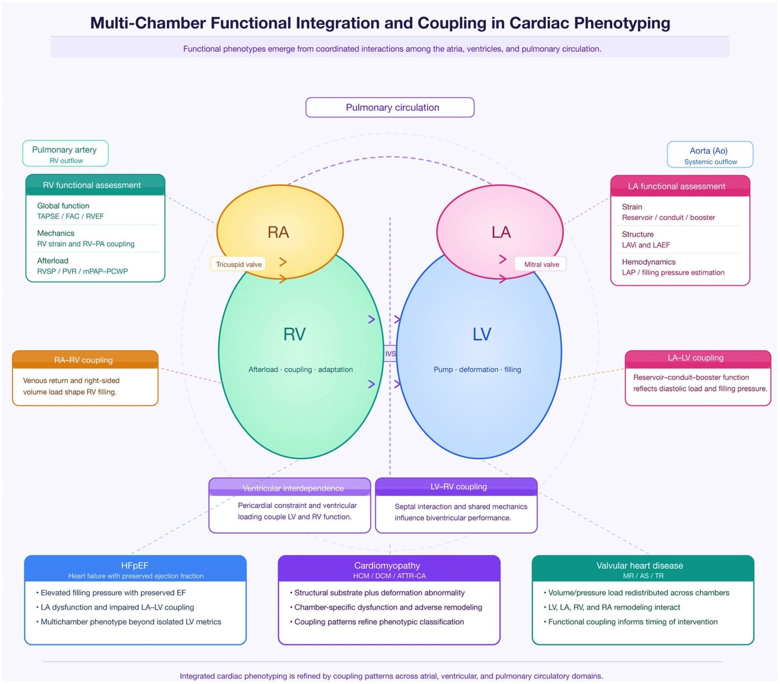
Heart failure cannot be fully understood as an isolated disorder of LV ejection. The syndrome reflects interactions among multiple cardiac chambers and the pulmonary and systemic circulation. As a result, increasingly sophisticated echocardiographic phenotyping is expanding beyond LV EF toward a multichamber assessment.
The right ventricle is particularly important. RV dysfunction may develop as a consequence of elevated left-sided filling pressures, pulmonary hypertension, intrinsic RV disease, ischemia, or ventricular interdependence. Conventional measures such as TAPSE, tissue Doppler S′, and fractional area change provide useful information, but each captures only selected aspects of RV performance.
RV global longitudinal strain offers a deformation-based measure that may identify subtle RV dysfunction. In HFpEF, impaired RV mechanics can be associated with pulmonary vascular abnormalities and adverse outcomes. The combination of RV strain, RV size, tricuspid regurgitation severity, estimated pulmonary artery pressure, and right atrial parameters may therefore provide a more complete picture of right-sided involvement.
The left atrium is equally important. Chronic elevation of LV filling pressures can produce atrial enlargement and progressive structural remodeling. However, atrial volume reflects cumulative remodeling and may not capture functional impairment at an early stage. Left atrial strain can characterize reservoir, conduit, and contractile function and may therefore provide additional information about ventricular–atrial coupling.
The interaction between LV and LA mechanics is particularly relevant in HFpEF. The stiff LV may require higher filling pressures to achieve adequate preload, while the left atrium may progressively lose its ability to buffer those pressure changes. During exercise, these abnormalities may become more pronounced, explaining why resting measurements can occasionally fail to capture the patient’s physiological limitation.
This has stimulated interest in exercise echocardiography and diastolic stress testing. Patients with apparently normal resting filling pressures may demonstrate abnormal increases during exertion. Combining stress hemodynamics with strain and chamber mechanics may therefore reveal latent dysfunction that is not apparent at rest.
Ventricular–arterial coupling represents another layer of complexity. The heart operates against an arterial system whose resistance and compliance influence myocardial workload. A preserved EF does not necessarily indicate efficient ventricular–arterial interaction. Patients with hypertension and HFpEF may maintain EF despite increased myocardial workload and altered geometry.
A comprehensive echocardiographic phenotype can therefore include LV GLS, LV volumes, LV mass, diastolic indices, LA volume and strain, RV strain and function, tricuspid regurgitation, pulmonary pressure estimates, and IVC characteristics. The challenge is to transform this large set of measurements into clinically interpretable information.
This is where computational methods may become increasingly valuable. Humans are excellent at recognizing meaningful patterns but may struggle to integrate dozens of partially correlated quantitative variables consistently. Machine learning can potentially identify combinations of measurements associated with specific outcomes or disease phenotypes.
However, computational complexity should not become an excuse for clinically opaque models. The objective should be interpretable augmentation of clinical reasoning rather than replacing it with unexplained scores. A useful multichamber model should ideally provide not only a risk estimate but also identify the dominant physiological abnormalities contributing to that estimate.
The future of echocardiographic heart failure phenotyping may therefore resemble a multidimensional map rather than a single classification label. Such a map could describe systolic mechanics, diastolic reserve, atrial function, RV–pulmonary coupling, chamber geometry, and hemodynamic burden.
This approach is especially relevant to HFpEF, where disease heterogeneity has historically limited the effectiveness of one-size-fits-all diagnostic frameworks. A richer imaging phenotype may help distinguish patients with predominantly myocardial dysfunction from those whose symptoms are driven primarily by pulmonary vascular disease, atrial dysfunction, valvular disease, obesity-related hemodynamic abnormalities, or other mechanisms.
Artificial Intelligence in Echocardiography: From Automated Measurements to Disease Phenotyping
Artificial intelligence is transforming cardiovascular imaging by changing how echocardiographic information can be acquired, processed, quantified, and interpreted. In conventional echocardiography, the clinician or sonographer must identify cardiac structures, select appropriate frames, trace borders, calculate volumes, estimate functional parameters, and interpret multiple measurements. Each step introduces potential variability.
AI can automate portions of this workflow. Modern deep-learning systems can perform image view recognition, chamber segmentation, border detection, volumetric calculation, EF estimation, and—in selected systems—strain analysis. The American Heart Association has highlighted applications of AI in echocardiography that include automated segmentation, volumetric analysis, EF calculation, valve assessment, myocardial deformation, and automated disease detection.
The more ambitious application is disease phenotyping. Instead of asking an algorithm to calculate EF, researchers are increasingly training models to recognize complex patterns associated with disease. This distinction is important. A measurement algorithm reproduces a human-defined parameter; a disease-classification model may discover combinations of features that are difficult to define explicitly.
HFpEF is an important example. Diagnostic assessment often requires integration of symptoms, natriuretic peptides, atrial and ventricular structure, Doppler measurements, and clinical context. AI models trained on echocardiographic videos have demonstrated the ability to identify HFpEF with promising discrimination in research cohorts. These findings suggest that apparently subtle motion patterns within routine echocardiographic images may contain diagnostic information that is difficult to extract through conventional measurements alone.
AI may also help address the problem of indeterminate cases. Current HFpEF diagnostic scores can leave a proportion of patients in an intermediate category. An imaging-based AI system could potentially provide an additional probability estimate, prioritize patients for further testing, or identify patients requiring exercise or invasive hemodynamic assessment.
However, impressive model performance does not automatically establish clinical utility. AI systems can perform extremely well in retrospective datasets while failing when transferred to different hospitals, ultrasound machines, patient populations, or acquisition protocols. Differences in prevalence, demographics, comorbidity burden, image quality, and labeling practices can substantially affect performance.
Generalizability is therefore one of the central challenges of cardiovascular AI. External validation across institutions, countries, ethnic groups, imaging platforms, and clinical environments is essential. Prospective evaluation is even more important because the ultimate question is whether AI improves clinical decisions and patient outcomes.
Interpretability is another major issue. A model that labels an echocardiogram as “high risk” without identifying relevant features may be difficult to trust. Explainable AI techniques may help by showing regions of the image that contributed to the prediction or by combining the prediction with interpretable measurements such as strain, chamber volume, and wall-motion abnormalities.
AI also introduces ethical and regulatory considerations. Training datasets may contain systematic biases. Algorithms may perform differently across demographic groups, and automated outputs can create false confidence if clinicians assume that machine-generated measurements are inherently objective.
The correct future model is therefore clinician-AI collaboration. AI should reduce repetitive measurement tasks, identify patterns requiring attention, improve consistency, and provide decision support while leaving clinical responsibility with appropriately trained professionals.
A particularly important development will be multimodal AI. Rather than analyzing echocardiographic images alone, future systems may integrate imaging, ECG, laboratory biomarkers, clinical records, genetics, wearable-device data, and longitudinal outcomes. Such systems could potentially create dynamic patient-specific cardiac phenotypes.
This possibility is scientifically exciting but should be approached cautiously. The more data an algorithm integrates, the more important transparency, validation, calibration, privacy protection, and clinical interpretability become. The future of AI in heart failure should therefore be judged not by algorithmic sophistication alone, but by whether it produces reproducible, equitable, clinically actionable improvements.
Integrating Strain, 3D Echocardiography, and Artificial Intelligence Into a Multidimensional Heart Failure Model
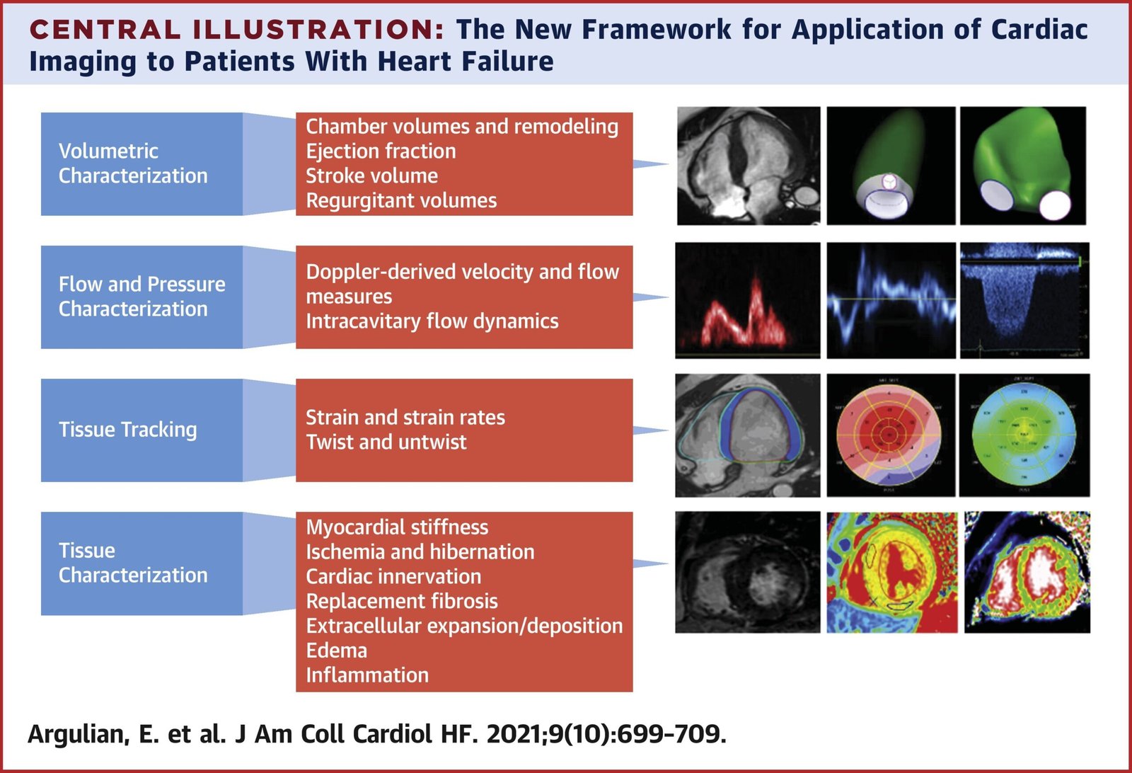
The greatest opportunity does not lie in any single technology. It lies in combining complementary technologies to construct a more complete representation of cardiac function. LVEF provides chamber-level systolic performance; strain describes myocardial deformation; 3D echocardiography characterizes chamber volumes and geometry; Doppler provides hemodynamic information; and AI can integrate large numbers of variables.
A multidimensional model could begin with conventional parameters. LVEF, LV end-diastolic and end-systolic volumes, wall thickness, LV mass, left atrial volume, RV dimensions, valvular abnormalities, and Doppler measurements provide the structural and hemodynamic foundation.
Strain would then add myocardial mechanics. LV GLS could quantify global longitudinal dysfunction, while segmental strain could identify heterogeneous patterns. RV strain and LA strain could provide additional information about multichamber involvement.
Three-dimensional imaging would contribute more accurate chamber geometry and volumetric measurements. In patients with remodeling, it may help distinguish dilated phenotypes from concentric remodeling and provide a more precise assessment of reverse remodeling.
AI could operate at several levels. At the simplest level, it could automate image quality control and measurement. At an intermediate level, it could combine measurements to estimate disease probability or prognosis. At the most advanced level, deep-learning models could analyze raw image sequences and discover latent imaging features not represented by conventional measurements.
This framework could potentially produce a patient-specific cardiac phenotype rather than a single diagnostic label. For example, a patient could demonstrate preserved LVEF but reduced GLS, increased LA volume, impaired LA strain, increased estimated filling pressure, and subtle RV dysfunction. Such a phenotype would be very different from another patient with the same EF but normal strain, normal atrial mechanics, and no evidence of elevated filling pressure.
The clinical advantage of such phenotyping is potentially substantial. It may identify disease earlier, improve risk stratification, guide additional testing, and help determine which patients require closer follow-up. It may also improve research by allowing investigators to enroll more biologically homogeneous populations.
However, multidimensional phenotyping creates a risk of overfitting and complexity. More measurements do not necessarily mean better medicine. Some variables may be redundant, poorly reproducible, or strongly dependent on loading conditions. The objective should therefore be parsimonious integration of validated parameters rather than indiscriminate accumulation of data.
Another challenge is defining clinically meaningful thresholds. While certain strain values are widely used in research, strain is not yet a universal binary diagnostic test for heart failure. Similarly, AI-derived probability scores require calibration and validation before they can be translated into clinical decision thresholds.
Longitudinal modeling may ultimately be more valuable than static phenotyping. Heart failure evolves over time, and serial imaging can reveal trajectories of ventricular remodeling, strain deterioration or improvement, atrial dysfunction, and treatment response. AI may be particularly useful in recognizing these trajectories because it can compare large numbers of parameters across repeated examinations.
Such a system could theoretically identify a patient whose LVEF has remained stable but whose myocardial strain has progressively deteriorated, or another patient whose EF has improved while residual abnormalities in strain and atrial mechanics persist. These trajectories may contain clinically relevant information that a single examination cannot provide.
The emerging model is therefore best understood as a transition from “measurement” to “phenotype,” and from “phenotype” to “trajectory.” The objective is not merely to describe what the heart looks like today but to understand where it is moving.
Clinical Applications: Early Detection, Prognosis, Treatment Response, and Precision Cardiology
The first major clinical application of advanced echocardiographic phenotyping is early recognition of myocardial dysfunction. Patients with hypertension, diabetes, obesity, cardiotoxic exposure, ischemic disease, or structural heart disease may develop myocardial abnormalities before conventional EF declines. Strain may identify subtle dysfunction, while 3D imaging can characterize remodeling and AI may integrate multiple weak signals into a stronger risk estimate.
The second application is prognosis. Numerous studies have linked abnormal GLS and other advanced echocardiographic measurements with mortality and hospitalization. Importantly, the prognostic value of these parameters may persist after accounting for conventional EF, supporting their role as complementary biomarkers.
In established heart failure, advanced imaging can also assist in treatment monitoring. Reverse remodeling may manifest as reductions in ventricular volumes, improvement in strain, changes in atrial function, and improvement in hemodynamic parameters. However, each parameter responds differently to therapy, and no single imaging marker should be assumed to represent treatment success.
The third application is patient selection for additional diagnostic testing. A patient with unexplained symptoms and preserved EF but abnormal strain and abnormal atrial mechanics may warrant more comprehensive evaluation for HFpEF, ischemic disease, infiltrative cardiomyopathy, valvular disease, or pulmonary hypertension depending on the clinical context.
Advanced imaging may also assist in differentiating apparently similar HF phenotypes. Patients with preserved EF can have markedly different pathophysiology. Identifying the dominant mechanism may become increasingly important as treatment becomes more phenotype-driven.
In cardio-oncology, strain has a particularly established conceptual role because myocardial dysfunction may develop before a conventional EF decline. Serial imaging may therefore support earlier recognition of treatment-associated cardiac dysfunction.
In valvular disease, 3D imaging can improve anatomical characterization while strain may reveal ventricular consequences of chronic pressure or volume overload. In cardiomyopathy, segmental deformation patterns may support etiological evaluation and guide subsequent CMR or genetic investigation.
AI may expand these applications by improving scalability. A major limitation of advanced echocardiography is that sophisticated analysis can be time-consuming and operator-dependent. Automated algorithms could make measurements available more consistently without requiring every laboratory to maintain highly specialized expertise.
AI-guided handheld echocardiography represents another potentially important development. If automated acquisition guidance and interpretation become sufficiently reliable, cardiac ultrasound could potentially move into primary care, emergency medicine, inpatient screening, and resource-limited environments. Recent studies demonstrate promising performance for AI-supported focused ultrasound and automated analysis, but broader validation remains essential.
Precision cardiology is the long-term objective. Instead of classifying patients only by EF, clinicians could potentially use integrated phenotypes to determine who needs further testing, who requires closer surveillance, and who may have a particular biological substrate.
Nevertheless, the distinction between prognostic association and treatment utility must remain explicit. A parameter can predict mortality without necessarily improving outcomes when incorporated into treatment decisions. Before advanced imaging biomarkers become treatment-selection tools, prospective interventional studies are needed to demonstrate that acting on the information changes clinical outcomes.
This is particularly important for AI. A model can achieve impressive AUC values and still provide little benefit if clinicians do not know what action to take based on its output. Clinical implementation therefore requires not only accurate prediction but also a defined decision pathway.
The most useful systems will likely be those that answer actionable questions: Should this patient undergo additional testing? Is there evidence of early myocardial dysfunction? Is the patient’s phenotype changing? Is the examination technically reliable? Does the imaging pattern suggest a particular etiology? Does the patient’s trajectory warrant intensified surveillance?
Advanced echocardiography will ultimately succeed when it becomes clinically actionable rather than merely technologically impressive.
Limitations, Standardization, Reproducibility, and Implementation Challenges
The enthusiasm surrounding strain, 3D echocardiography, and AI must be balanced by recognition of important limitations. Advanced imaging can increase information density, but more information does not automatically mean more accurate diagnosis or better patient outcomes.
Strain is affected by image quality, frame rate, software algorithms, vendor differences, loading conditions, and acquisition technique. Differences between platforms can make direct comparison of absolute values problematic. This creates challenges for multicenter research, longitudinal monitoring, and universal threshold development.
Three-dimensional echocardiography has its own limitations. Adequate acoustic windows are required, and temporal and spatial resolution can be inferior to selected two-dimensional acquisitions. Stitching artifacts may occur during multibeat acquisition, particularly in patients with arrhythmias or respiratory instability. These issues may reduce reliability in some clinical settings.
AI introduces a different class of challenges. Data quality is fundamental. If training data contain inaccurate labels, systematic acquisition differences, or demographic imbalance, the resulting model may reproduce or amplify those limitations.
External validation is therefore essential. A model developed at a tertiary academic center may not perform identically in a community hospital, a different country, or a population with different disease prevalence. The model must be evaluated across diverse settings before broad clinical adoption.
There is also the problem of dataset shift. Ultrasound machines are updated, acquisition protocols change, software evolves, and patient populations change. An algorithm that performs well today may require continuous monitoring to ensure that its performance remains acceptable.
Clinical interpretability is another challenge. Clinicians need to understand when an automated output is reliable and when it should be ignored. Systems should ideally provide confidence estimates, quality indicators, and interpretable supporting features.
Automation bias must also be considered. If clinicians assume that a computer-generated measurement is inherently more objective than a human measurement, they may fail to recognize obvious errors. AI should therefore function as decision support rather than an unquestioned authority.
Regulatory and ethical issues are equally important. AI systems may influence diagnosis and clinical decisions, meaning that governance, transparency, cybersecurity, data privacy, and accountability must be considered from the beginning.
Another challenge is workflow integration. An algorithm that requires exporting images to a separate platform, waiting for processing, and manually transferring results into the electronic record may create additional burden rather than reduce it.
Cost-effectiveness must also be established. Advanced imaging and AI technologies require investment in equipment, software, training, infrastructure, and quality assurance. Their adoption should therefore be justified by demonstrable improvements in diagnostic accuracy, efficiency, outcomes, or resource utilization.
The most important methodological principle is that technical validation is not enough. Future studies should progress from retrospective accuracy studies to prospective multicenter validation and ultimately to clinical utility trials. The relevant endpoint should increasingly be whether use of the technology changes management appropriately and improves patient outcomes.
Standardization efforts will therefore be critical. Common acquisition protocols, reference populations, reporting frameworks, quality-control procedures, and transparent AI evaluation standards will be necessary to transform promising technologies into dependable clinical tools.
The future of advanced cardiac imaging depends not on eliminating uncertainty but on quantifying it. A clinically mature system should know when it is confident, when image quality is inadequate, and when additional testing is required.
Future Directions: Toward AI-Enabled Digital Cardiac Phenotypes
The next phase of echocardiographic innovation will likely involve the transition from isolated measurements to dynamic digital cardiac phenotypes. Instead of generating a conventional report containing dozens of unrelated numbers, future systems may integrate structural, mechanical, hemodynamic, and temporal information into a coherent representation of the patient’s cardiovascular state.
A digital phenotype could incorporate LVEF, three-dimensional ventricular volumes, GLS, segmental strain, RV mechanics, atrial strain, diastolic indices, pulmonary pressure estimates, valvular characteristics, ECG features, biomarkers, and longitudinal clinical data.
AI is uniquely positioned to integrate these heterogeneous data types. Deep-learning models can process image sequences, while multimodal architectures can combine imaging with structured clinical information. Such systems may identify subtle patterns that are difficult to encode using traditional rule-based algorithms.
One potential future application is automated detection of preclinical heart failure. Rather than waiting for symptomatic disease, an AI system could identify a pattern of early myocardial dysfunction and estimate the probability of progression. This would be particularly relevant to high-risk populations.
Another possibility is personalized trajectory prediction. Serial echocardiograms could be analyzed to estimate whether a patient is undergoing favorable reverse remodeling, stable disease, or progressive deterioration. The trajectory itself may become a clinically meaningful biomarker.
Digital twins represent a more ambitious concept. A computational model of a patient’s cardiac mechanics could theoretically simulate changes in ventricular loading, myocardial function, and treatment response. Although this remains largely a research concept, advances in 3D imaging, strain, computational modeling, and AI may gradually make increasingly sophisticated patient-specific simulations possible.
The integration of wearable and remote monitoring data could further expand this model. Heart rate, activity, respiratory rate, blood pressure, weight trends, and other longitudinal signals might be combined with periodic imaging to create a dynamic cardiovascular profile.
However, the future should not be driven by technology for its own sake. The central question must remain whether these systems improve patient care. An algorithm that produces a more complex phenotype without improving clinical decisions has limited practical value.
Future research should therefore focus on prospective outcome studies, external validation, equity, interpretability, cost-effectiveness, and clinical workflow integration. Models should be evaluated across diverse populations and healthcare systems, with transparent reporting of performance and failure modes.
The field also needs better definitions of clinically meaningful change. For strain, 3D volumes, and AI-derived scores, the difference between biological change and measurement noise must be quantified. This is essential before these parameters can be confidently used for treatment monitoring.
Another important direction is explainable multimodal AI. Rather than producing only a probability score, future systems should ideally identify the dominant imaging features contributing to the result. For example, a model might indicate that its prediction is driven primarily by reduced GLS, increased LA volume, abnormal atrial strain, and elevated filling-pressure surrogates.
This approach could make AI more clinically acceptable because it aligns computational predictions with recognizable cardiovascular physiology.
The ultimate goal is therefore not to replace echocardiography or clinicians but to make cardiac imaging more quantitative, reproducible, scalable, and biologically informative. The convergence of myocardial strain, 3D echocardiography, and AI offers a pathway toward this objective.
Conclusions
Heart failure phenotyping is undergoing a fundamental transition. LVEF remains an essential clinical parameter, but it cannot independently describe the complexity of myocardial dysfunction. Patients with similar EF values may have profoundly different mechanical, structural, hemodynamic, and prognostic profiles.
Myocardial strain provides a window into deformation and can reveal subclinical dysfunction that may remain hidden when assessment relies primarily on EF. GLS has demonstrated prognostic value across multiple cardiovascular conditions and appears particularly relevant in patients with preserved or mildly reduced EF.
Three-dimensional echocardiography complements this information by providing more comprehensive assessment of ventricular geometry and volumes. Its ability to characterize remodeling and chamber structure may become increasingly important as heart failure phenotyping moves beyond a single EF measurement.
Artificial intelligence adds another dimension by enabling automated quantification and recognition of complex imaging patterns. Emerging studies demonstrate that AI can detect heart failure phenotypes from echocardiographic data, including HFpEF, with promising performance. Nevertheless, generalizability, transparency, bias, regulatory oversight, and clinical utility remain major challenges.
The most promising future is therefore an integrated model in which conventional echocardiography, myocardial deformation, three-dimensional structural analysis, and AI-based pattern recognition operate together. Such an approach could move heart failure imaging from static classification toward early detection, multidimensional phenotyping, and longitudinal trajectory assessment.
The central principle should remain clinically grounded: advanced imaging should not replace established diagnostic frameworks but should enrich them. The future of heart failure assessment is unlikely to be defined by a single superior parameter. Instead, it will depend on how effectively multiple complementary signals can be integrated into a reliable, interpretable, and actionable representation of cardiac biology.
Beyond ejection fraction, the emerging question is no longer simply how much blood the heart ejects. It is how the myocardium deforms, how the chambers remodel, how pressures interact, how the phenotype evolves, and how intelligently these signals can be combined to identify disease earlier and characterize it more precisely.