



1. Hepatic Encephalopathy as a Multisystem Complication of Cirrhosis
Hepatic encephalopathy (HE) is one of the most clinically important complications of advanced liver disease, yet its biological complexity is frequently underestimated. Historically, HE was conceptualized primarily as a consequence of hepatic failure to detoxify ammonia. This model remains fundamentally important, but contemporary evidence indicates that ammonia alone cannot explain the remarkable variability in neurological manifestations among patients with apparently similar degrees of cirrhosis or similar circulating ammonia concentrations.
HE should instead be regarded as a dynamic neurological syndrome emerging from the interaction between hepatic insufficiency, portal-systemic shunting, altered nitrogen metabolism, systemic inflammation, intestinal dysbiosis, intestinal barrier dysfunction, skeletal-muscle wasting, electrolyte disturbances, medications, and cerebral susceptibility. Recent literature increasingly describes HE as a gut–liver–brain or even gut–liver–muscle–brain disorder rather than an isolated consequence of hepatic detoxification failure.
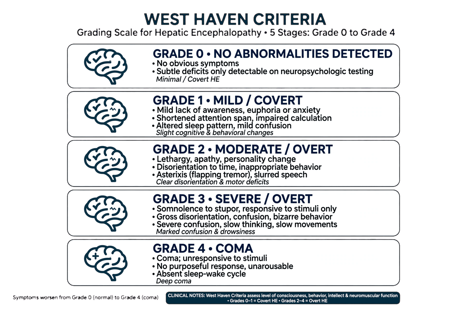
The clinical spectrum is broad. At one end are covert forms, including minimal and grade I HE, in which conventional bedside examination may appear almost normal despite measurable deficits in attention, psychomotor speed, executive function, visuospatial processing, and reaction time. At the other end is overt HE, which can progress from disorientation and asterixis to profound somnolence, coma, and death.
This spectrum creates an important clinical challenge. Overt HE is relatively recognizable, but covert HE can remain undiagnosed for prolonged periods. Patients may complain only of impaired concentration, sleep disturbance, reduced work performance, irritability, slowed decision-making, or difficulty performing complex tasks. These abnormalities may have profound consequences for driving, employment, medication adherence, falls, caregiver burden, and quality of life.
The relationship between HE and cirrhosis is also bidirectional. Advanced liver disease increases susceptibility to HE, while recurrent HE is associated with hospitalization, functional decline, frailty, and mortality. Each episode may represent not only a neurological complication but also a marker of worsening hepatic reserve and systemic vulnerability.
The traditional “ammonia-only” model therefore becomes inadequate when attempting to explain why some patients with cirrhosis develop severe neurological dysfunction while others with similar ammonia levels remain relatively asymptomatic.
A more useful conceptual model is a systems framework.
The failing liver reduces ammonia clearance and alters metabolism of multiple circulating substances.
Portal hypertension creates collateral circulation that bypasses hepatic detoxification.
The gut produces ammonia and other potentially neuroactive metabolites.
The intestinal barrier becomes dysfunctional.
Microbial composition and metabolic function change.
Systemic inflammation increases blood–brain barrier vulnerability.
Skeletal muscle loss reduces an important extrahepatic site of ammonia disposal.
The brain responds to ammonia and inflammatory signaling through astrocytic swelling, altered neurotransmission, oxidative stress, and impaired energy metabolism.
The resulting neurological syndrome is therefore the product of multiple interacting biological processes.
This framework also explains why therapeutic approaches that only reduce ammonia may improve HE without completely normalizing cognition in every patient.
It explains why rifaximin can provide benefit through mechanisms extending beyond simple suppression of ammonia-producing bacteria.
It explains why microbiome manipulation has become an area of intense research.
And it explains why sarcopenia is increasingly recognized as an important modifier of HE risk.
The modern challenge is therefore not to replace the ammonia hypothesis but to place it within a broader network.
Ammonia remains a major neurotoxin.
It is not, however, the entire disease.
This distinction has practical consequences.
Serum ammonia should not be interpreted as a stand-alone diagnostic test.
A patient with cirrhosis and confusion requires clinical evaluation for HE, but also for infection, bleeding, electrolyte disturbances, renal dysfunction, medications, intoxication, hypoglycemia, intracranial disease, and other causes of altered mental status.
The American Association for the Study of Liver Diseases emphasizes that HE remains a clinical diagnosis and that ammonia concentration does not reliably correlate with HE grade.
The emerging paradigm therefore shifts the central question from:
“How high is the ammonia?”
to: “Why has this patient’s brain become dysfunctional in this particular physiological context?”
That question is more difficult, but it is also more clinically useful.
The future of HE management will likely depend on identifying the combination of hepatic, microbial, inflammatory, metabolic, muscular, and neurological factors that define an individual’s vulnerability.
This is the foundation of precision hepatology.
2. From Ammonia to Neurotoxicity: Molecular Mechanisms of Brain Dysfunction
Ammonia remains the central metabolic toxin in hepatic encephalopathy, and any modern model that minimizes its importance would be scientifically misleading. The critical evolution has instead been the recognition that ammonia acts through multiple downstream pathways and that its neurological effects depend heavily on the biological environment in which it accumulates.
Ammonia is generated throughout the body but the gastrointestinal tract represents a major source. Intestinal bacteria metabolize nitrogen-containing substrates, while enterocytes and other tissues contribute additional ammonia through amino-acid metabolism. Under normal conditions, the liver removes ammonia through the urea cycle and glutamine synthesis. In cirrhosis, hepatic detoxification becomes impaired and portal-systemic shunting allows ammonia to bypass hepatocytes.
Skeletal muscle becomes an increasingly important alternative site of ammonia disposal. Muscle converts ammonia into glutamine through glutamine synthetase. Consequently, severe sarcopenia can reduce systemic ammonia buffering capacity. This creates an important biological connection between malnutrition, frailty, and HE.
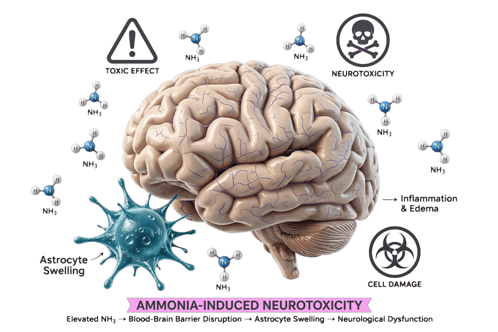
Once ammonia reaches the brain, astrocytes become a major target.
Astrocytes contain glutamine synthetase and convert ammonia plus glutamate into glutamine. Excessive intracellular glutamine can increase osmotic stress and contribute to astrocyte swelling. In severe acute liver failure, this mechanism is strongly associated with cerebral edema and intracranial hypertension. In chronic cirrhosis, the process is generally more complex and may involve chronic astrocytic adaptation rather than simple acute swelling.
Ammonia also alters mitochondrial function and generates oxidative and nitrosative stress. Reactive oxygen and nitrogen species can impair cellular signaling and energy metabolism, while inflammatory pathways may amplify neuronal dysfunction.
Neurotransmission is affected as well.
HE has been associated with alterations in GABAergic, glutamatergic, dopaminergic, serotonergic, and other neurotransmitter systems. The traditional “false neurotransmitter” and benzodiazepine-like theories are now viewed as components of a larger network rather than complete explanations.
Ammonia can also alter cerebral blood flow and neurovascular signaling.
The blood–brain barrier itself becomes functionally vulnerable in the context of systemic inflammation, oxidative stress, and cirrhosis-associated endothelial dysfunction.
This creates an important interaction.
Ammonia may prime the brain.
Inflammation may amplify the response.
Infection may abruptly worsen neurological dysfunction.
Electrolyte abnormalities may lower the threshold for cognitive impairment.
Sedative medications may further suppress consciousness.
This explains why HE often behaves as a threshold phenomenon.
A patient may have chronic compensated metabolic abnormalities without overt symptoms until an additional insult—such as infection, gastrointestinal bleeding, constipation, dehydration, renal failure, or sedative exposure—pushes cerebral function beyond a critical threshold.
Recent work has expanded this model even further by identifying microbial neurotoxins other than ammonia. A 2025 Nature Medicine study identified increased phenylalanine decarboxylase genes, particularly from Ruminococcus gnavus, in patients with cirrhosis and HE. The investigators linked microbial production of phenylethylamine to neurological abnormalities in experimental models and found that higher baseline phenylethylamine was associated with markedly increased risk of HE after TIPS.
This is an important conceptual development.
It does not mean that ammonia has become irrelevant.
It means that the intestine may deliver a complex mixture of neuroactive metabolites to the circulation, with ammonia representing one major component of a much larger chemical ecosystem.
The brain is therefore exposed not simply to one toxin but to a changing metabolic environment.
This may explain why patients with similar ammonia concentrations can have dramatically different neurological phenotypes.
The clinical implications are substantial.
A future biomarker panel may need to capture not only ammonia but also inflammatory mediators, microbial metabolites, bile-acid signaling, markers of astrocytic stress, and measures of systemic metabolic vulnerability.
Therapeutically, the goal may shift from simply lowering ammonia to modifying the entire metabolic environment that determines how the brain responds to ammonia.
This represents a major evolution in the pathophysiological model of HE.
The ammonia hypothesis remains the foundation.
But the modern version is no longer:
ammonia → brain dysfunction.
It is closer to:
gut nitrogen metabolism + impaired hepatic clearance + portal shunting + muscle dysfunction + systemic inflammation + microbial metabolites + astrocytic vulnerability → neurological dysfunction.
That model better reflects contemporary evidence and creates a broader therapeutic landscape.
3. The Gut–Liver–Brain Axis: Microbiome, Intestinal Barrier, and Microbial Neurotoxins
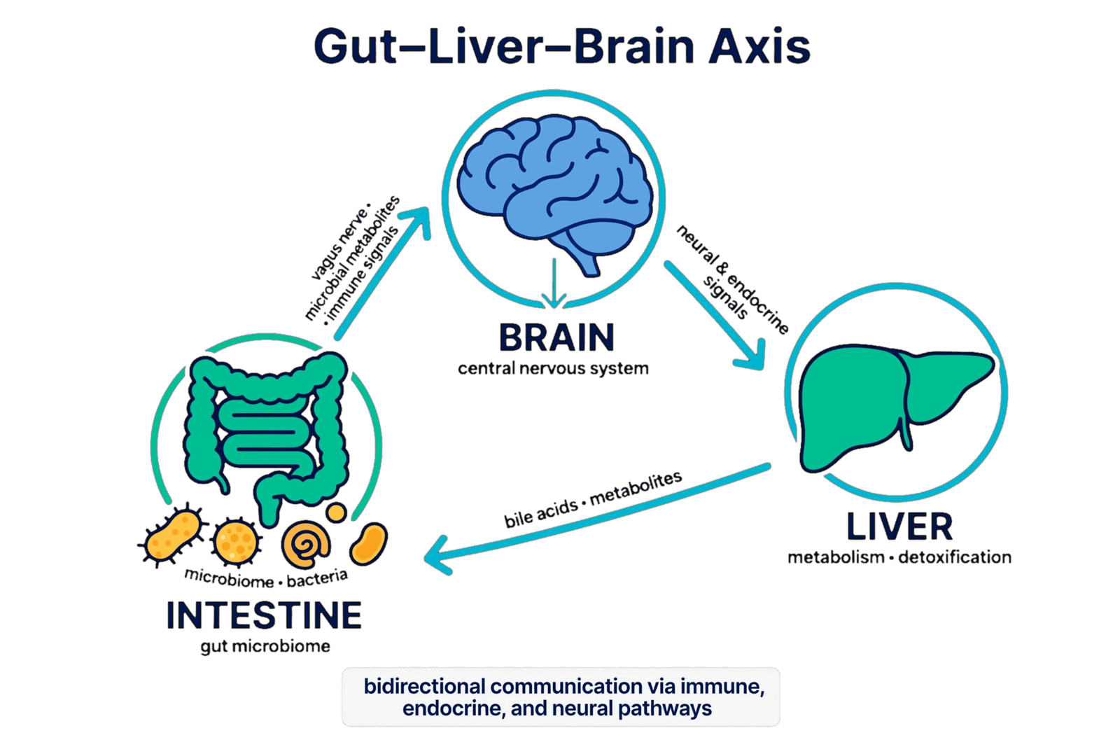
The gut–liver–brain axis has become one of the most important conceptual frameworks in modern hepatic encephalopathy research. It recognizes that the intestine is not merely a passive source of ammonia but an active metabolic and immunological organ capable of influencing hepatic inflammation, systemic metabolism, and brain function.
Cirrhosis is associated with profound changes in intestinal microbial ecology. Reduced microbial diversity, expansion of potentially pathogenic organisms, loss of beneficial taxa, altered microbial metabolism, and impaired intestinal barrier integrity can occur as liver disease progresses.
These changes matter because the microbiome produces far more than ammonia.
Microbial metabolism generates short-chain fatty acids, bile-acid derivatives, tryptophan metabolites, phenolic compounds, indoles, neurotransmitter-related molecules, and other metabolites capable of influencing immune and neurological pathways.
The intestinal barrier is equally important.
A healthy intestinal epithelium restricts translocation of bacteria and microbial products. In cirrhosis, portal hypertension, altered bile acids, inflammation, and dysbiosis can compromise barrier integrity. Increased permeability allows microbial-associated molecular patterns such as lipopolysaccharide to enter the portal and systemic circulation.
These signals activate innate immune pathways.
Kupffer cells and systemic immune cells respond with inflammatory mediators.
The liver becomes more inflamed.
The systemic inflammatory state becomes more pronounced.
The brain is subsequently exposed to a milieu that can increase neuroinflammation and alter blood–brain barrier function.
This creates a biological feedback loop:
cirrhosis → intestinal dysbiosis → barrier dysfunction → microbial translocation → systemic inflammation → brain vulnerability → HE.
At the same time, hepatic dysfunction alters bile-acid metabolism and intestinal conditions, which can further reshape microbial communities.
The axis is therefore bidirectional.
The gut affects the liver.
The liver affects the gut.
Both affect the brain.
The concept has gained strong experimental support. Reviews of the microbiota–gut–liver–brain axis describe interactions involving immune, metabolic, neuroendocrine, and vagal pathways, while recent mechanistic studies have identified specific microbial metabolites with potential neurological effects.
The 2025 Nature Medicine study involving Ruminococcus gnavus is particularly provocative because it moves beyond broad descriptions of “dysbiosis.” The investigators identified a specific microbial enzymatic pathway capable of generating phenylethylamine and linked this metabolite to neurological abnormalities in cirrhotic experimental models.
This type of work represents the next generation of microbiome research.
Rather than asking whether the microbiome is “good” or “bad,” researchers can ask:
Which organism?
Which gene?
Which metabolic pathway?
Which metabolite?
Which host receptor?
Which neurological phenotype?
This is much closer to precision medicine.
However, caution is necessary.
Microbiome studies frequently identify associations rather than causation.
Microbial composition varies according to geography, diet, antibiotics, proton-pump inhibitors, hospitalization, etiology of cirrhosis, alcohol exposure, stool transit, and many other variables.
Therefore, identifying an altered microbial taxon does not automatically establish that the organism causes HE.
This is one reason why interventions based on microbiome sequencing remain investigational.
The clinical microbiome is also highly dynamic.
A patient receiving lactulose may have a different microbial profile from one receiving rifaximin.
Antibiotics for spontaneous bacterial peritonitis can dramatically alter intestinal ecology.
FMT may produce substantial changes in microbial diversity, but whether those changes are durable and universally beneficial remains uncertain.
The most promising future approach may therefore be functional microbiomics.
Instead of asking only “which bacteria are present?”, clinicians may eventually evaluate what metabolic functions the microbial community is performing.
Metagenomics, metatranscriptomics, metabolomics, and host immune profiling could theoretically identify a patient-specific “HE signature.”
This could enable targeted interventions.
For example, one patient might have predominant ammonia production.
Another might have inflammatory microbial translocation.
Another might have excessive production of neuroactive aromatic metabolites.
Another might have profound sarcopenia and impaired ammonia disposal.
These patients might require different therapeutic strategies even though they share the same clinical diagnosis.
This is the essence of the gut–liver–brain paradigm.
HE may eventually be classified not only by clinical grade but by biological phenotype.
That possibility could transform treatment development.
Instead of developing another nonspecific ammonia-lowering drug, researchers could target a microbial enzyme, intestinal barrier pathway, bile-acid receptor, neuroinflammatory signal, or specific metabolite.
The gut–liver–brain axis therefore provides both a mechanistic explanation and a therapeutic roadmap.
The challenge now is to translate compelling mechanistic science into reproducible human clinical benefit.
4. Neuroinflammation, Astrocyte Dysfunction, Sarcopenia, and the Brain’s Metabolic Vulnerability
The neurological phenotype of hepatic encephalopathy cannot be explained by ammonia concentration alone because the brain’s response to ammonia depends strongly on inflammatory, metabolic, and structural conditions.
Astrocytes occupy a central position in this process.
These glial cells maintain extracellular neurotransmitter concentrations, regulate potassium, participate in energy metabolism, maintain the blood–brain barrier, and detoxify ammonia through glutamine synthesis.
When ammonia concentrations increase chronically, astrocytes undergo biochemical adaptation. Excess glutamine, altered mitochondrial metabolism, oxidative stress, and changes in intracellular signaling can compromise astrocytic function.
This may produce subtle cognitive dysfunction before overt neurological abnormalities become obvious.
Neuroinflammation adds another layer.
Cirrhosis is increasingly recognized as a systemic inflammatory disease, particularly during acute decompensation. Bacterial translocation, endotoxemia, infection, and inflammatory cytokines can activate microglia and alter neuronal signaling.
The interaction between ammonia and inflammation appears particularly important.
Ammonia can increase susceptibility to inflammatory injury.
Inflammation can increase the brain’s sensitivity to ammonia.
The combined effect may be substantially greater than either process alone.
This helps explain why infection can precipitate dramatic HE deterioration without necessarily producing an equivalent increase in serum ammonia.
Sarcopenia is another underappreciated determinant.
Skeletal muscle is an important site of ammonia detoxification through glutamine synthesis. When muscle mass decreases, the body’s capacity to buffer circulating ammonia is reduced.
Cirrhosis itself promotes muscle wasting through inadequate nutrition, hormonal alterations, inflammation, physical inactivity, and metabolic dysfunction.
This produces a vicious cycle:
cirrhosis → sarcopenia → reduced ammonia clearance → increased brain exposure → HE → reduced mobility and nutritional intake → further sarcopenia.
The implication is clinically important.
Nutrition and physical rehabilitation should not be regarded as secondary issues in HE management.
Adequate protein intake is generally encouraged rather than routinely restricted, because excessive protein restriction can worsen malnutrition and sarcopenia.
The modern nutritional strategy therefore focuses on sufficient energy, adequate protein, appropriate meal distribution, and prevention of prolonged fasting.
This represents another historical shift.
For decades, patients with HE were sometimes placed on restrictive protein diets because protein was assumed to drive ammonia production. Contemporary approaches recognize that chronic protein restriction can worsen muscle wasting and ultimately impair ammonia disposal.
Plant and dairy protein sources may be useful in some patients, but nutrition should be individualized.
Sleep disturbance also interacts with neurological vulnerability.
Patients with cirrhosis frequently experience abnormal sleep–wake cycles, daytime somnolence, insomnia, and circadian disruption. These symptoms can overlap with HE and make clinical assessment more difficult.
Electrolyte abnormalities are equally important.
Hyponatremia, hypokalemia, metabolic alkalosis, and renal dysfunction can increase susceptibility to neurological deterioration.
Sedative medications, particularly benzodiazepines and other central nervous system depressants, can further impair cognition.
The result is a complex threshold model.
The brain may tolerate a certain degree of hyperammonemia when systemic inflammation is low, hydration is stable, electrolytes are normal, and muscle mass is preserved.
The same ammonia concentration may produce severe dysfunction when infection, inflammation, renal dysfunction, hyponatremia, sarcopenia, and sedative exposure coexist.
This explains the imperfect relationship between serum ammonia and HE grade.
The AASLD emphasizes that ammonia concentrations do not consistently correlate with severity and that altered mental status in cirrhosis should not automatically be attributed to HE.
This broader model also explains why therapies that influence multiple biological pathways may be beneficial.
Lactulose changes intestinal metabolism and stool transit.
Rifaximin alters bacterial function.
Nutritional therapy preserves skeletal-muscle ammonia disposal.
Treating infection reduces inflammatory signaling.
Correcting electrolyte abnormalities restores neuronal stability.
Managing portal hypertension can alter shunting.
Liver transplantation addresses the underlying hepatic failure.
Thus, effective HE management is inherently multimodal.
A purely neurological approach is insufficient.
A purely hepatic approach is insufficient.
A purely microbiome approach is insufficient.
The syndrome emerges from the interaction of all these systems.
The future of HE care will therefore require closer integration of hepatology, nutrition, neurology, microbiology, rehabilitation, and transplant medicine.
The gut–liver–brain model is increasingly becoming a gut–liver–muscle–brain model.
That expansion is not merely semantic.
It identifies sarcopenia as a potentially modifiable component of neurological risk and emphasizes that preserving muscle may be as important as reducing ammonia production.
This is one of the most clinically actionable consequences of the modern pathophysiological framework.
5. Clinical Phenotypes: From Minimal and Covert HE to Overt and Recurrent Encephalopathy
Hepatic encephalopathy exists on a continuum rather than as a binary state.
The traditional West Haven framework categorizes overt HE from grade I through IV, ranging from subtle changes in attention and behavior to coma. However, clinically meaningful neurological impairment can occur before obvious disorientation develops.
This is the domain of covert hepatic encephalopathy.
Covert HE encompasses minimal HE and grade I abnormalities that may not be apparent during routine clinical examination but can be detected using specialized psychometric or neurophysiological testing.
These patients may appear normal during a brief clinical encounter.
Yet they may have impaired reaction time, attention, working memory, executive function, visuospatial processing, or psychomotor speed.
The consequences can be significant.
A patient who has subtle cognitive impairment may have difficulty driving safely.
Another may make medication errors.
Another may struggle with complex employment tasks.
Another may experience falls.
Family members may notice personality or sleep changes before clinicians recognize neurological disease.
Recent literature emphasizes that covert HE is strongly associated with quality-of-life impairment, progression to overt HE, and adverse outcomes.
This makes early identification clinically important.
However, routine screening is challenging.
Psychometric tests require time, education-adjusted interpretation, and appropriate normative data.
Cultural and language differences can influence performance.
Fatigue, depression, anxiety, visual impairment, and low educational attainment can produce false-positive results.
Therefore, no single cognitive test should be interpreted without context.
Point-of-care tools such as the Stroop test and animal naming test have gained interest because they can be performed relatively quickly and may identify cognitive impairment in outpatient settings. Recent reviews describe these tools as part of a broader diagnostic strategy for covert HE.
Overt HE is more clinically obvious.
Patients may demonstrate disorientation, confusion, inappropriate behavior, somnolence, asterixis, impaired consciousness, or coma.
Asterixis is characteristic but not specific.
It represents negative myoclonus and can occur in other metabolic encephalopathies.
Recurrent HE is particularly important because each episode increases healthcare utilization and often signals advanced disease.
The clinical challenge is not simply treating the current episode but preventing recurrence.
This requires identifying the precipitating factor, optimizing lactulose adherence and dosing, considering rifaximin in appropriate patients, correcting nutritional deficits, and reassessing liver disease severity and transplant candidacy.
An important distinction should be made between spontaneous and precipitated HE.
In many patients, HE is triggered by a reversible event.
Common precipitants include infection, gastrointestinal bleeding, constipation, dehydration, excessive diuresis, renal dysfunction, electrolyte abnormalities, sedative medications, and excessive dietary or protein changes.
Failure to identify the precipitant increases recurrence risk.
Another clinically important phenotype is HE associated with large spontaneous portosystemic shunts.
These patients may have relatively preserved hepatic synthetic function but recurrent or refractory encephalopathy because substantial portal blood bypasses hepatic detoxification.
In selected patients, shunt embolization can be considered.
TIPS-associated HE represents another special phenotype.
TIPS improves portal hypertension but increases portal-systemic shunting and may precipitate HE in susceptible patients.
The recent identification of phenylethylamine as a potential predictor after TIPS is an example of how emerging biomarkers could eventually improve risk stratification. In the 2025 mechanistic study, elevated baseline phenylethylamine was associated with approximately sevenfold higher risk of HE after intrahepatic portosystemic shunt procedures.
However, this remains a research finding rather than a validated routine clinical test.
This distinction is crucial.
A biomarker can be biologically fascinating without yet being clinically actionable.
Precision diagnosis therefore requires understanding the stage and context of HE rather than simply assigning a numerical grade.
The future may combine:
clinical West Haven assessment,
cognitive testing,
functional assessment,
ammonia and metabolic measurements,
inflammatory biomarkers,
microbiome signatures,
neuroimaging,
and digital cognitive monitoring.
Such multimodal assessment could identify patients at risk before overt HE develops.
The clinical objective should therefore evolve from treating coma to preventing neurological decline.
Covert HE should not be dismissed as “mild” simply because the patient looks normal.
It represents measurable brain dysfunction occurring before the neurological reserve has been completely exhausted.
Recognizing this window could allow intervention before recurrent hospitalization and overt encephalopathy occur.
6. Precision Diagnosis: Why Ammonia Alone Is Not Enough
One of the most persistent misconceptions in hepatic encephalopathy is that serum ammonia can establish or exclude the diagnosis.
It cannot.
Ammonia is mechanistically central, but its diagnostic performance is limited.
The AASLD explicitly notes that ammonia levels do not consistently correlate with HE severity and that normal and elevated levels may occur across different West Haven grades.
This creates an important distinction:
ammonia is a pathophysiological biomarker, not a stand-alone clinical diagnosis.
Several factors explain this limitation.
First, ammonia exists in equilibrium between NH3 and NH4+, and measured concentration is affected by sample handling, processing time, temperature, tourniquet use, and other preanalytical factors.
Second, blood ammonia does not necessarily reflect brain ammonia exposure.
Third, cerebral vulnerability varies among patients.
Fourth, inflammation, sarcopenia, renal dysfunction, and portal-systemic shunting modify the relationship between circulating ammonia and neurological manifestations.
The correct diagnostic approach begins with the clinical phenotype.
A patient with cirrhosis and a characteristic pattern of altered attention, behavior, consciousness, or motor findings may have HE.
But alternative diagnoses must be actively excluded.
This is especially important because cirrhosis is associated with multiple causes of altered mental status.
Hypoglycemia.
Hyperglycemia.
Hyponatremia.
Uremia.
Sepsis.
Intracranial hemorrhage.
Stroke.
Seizures.
Drug toxicity.
Alcohol intoxication or withdrawal.
Wernicke encephalopathy.
Hypercapnia.
Sedative accumulation.
All may mimic HE.
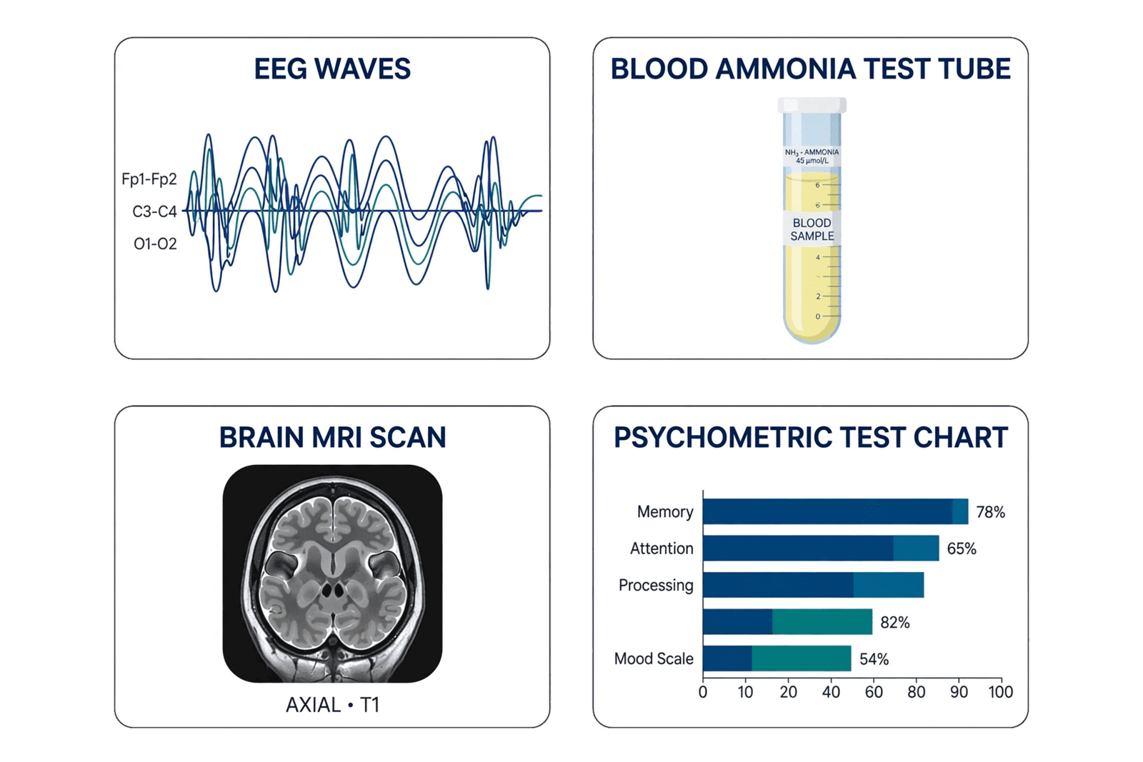
Brain imaging should therefore be considered when the presentation is atypical, focal neurological deficits are present, trauma is suspected, or the diagnosis remains uncertain.
Electroencephalography may be useful when nonconvulsive seizures are suspected.
For covert HE, psychometric and neurophysiological testing becomes more important.
The Psychometric Hepatic Encephalopathy Score remains a traditional tool, while newer point-of-care approaches such as Stroop-based testing can potentially increase accessibility.
The problem is standardization.
Different populations have different educational backgrounds, languages, age distributions, and cultural norms.
A precision diagnostic system must therefore interpret test performance against appropriate reference populations.
Biomarkers are an attractive solution.
Potential candidates include inflammatory cytokines, metabolomic signatures, microbial metabolites, bile-acid profiles, markers of astrocytic injury, and neuroimaging features.
But most remain investigational.
The 2025 literature emphasizes inadequate validated biomarkers as a major barrier to implementation of precision HE diagnosis. (PubMed)
Neuroimaging provides another potential dimension.
Magnetic resonance imaging can demonstrate abnormalities associated with chronic liver disease, including changes related to manganese deposition and alterations in brain metabolism or connectivity.
However, imaging is not currently a replacement for clinical diagnosis.
The future likely belongs to multimodal diagnosis.
Imagine a patient with cirrhosis who presents with subtle cognitive decline.
Rather than relying on ammonia alone, the clinician could combine:
clinical examination,
digital cognitive testing,
psychometric assessment,
ammonia,
renal and electrolyte status,
inflammatory biomarkers,
microbial metabolite profiles,
and imaging when indicated.
An AI model could potentially integrate these variables to estimate the probability of covert or overt HE and predict short-term deterioration.
However, this technology must be validated prospectively.
An algorithm that performs well in one tertiary center may fail in another population because microbiome composition, disease etiology, socioeconomic characteristics, and treatment patterns differ.
Precision medicine therefore requires not only sophisticated technology but rigorous external validation.
The most important current improvement, however, does not require AI.
It is simply abandoning ammonia-only reasoning.
A normal ammonia level should not automatically exclude HE when clinical suspicion is strong.
A high ammonia level should not automatically prove that altered mental status is due to HE.
Instead:
clinical phenotype + exclusion of mimics + cirrhosis context + supportive biomarkers = rational diagnosis.
This is the practical meaning of precision diagnosis in HE.
The goal is not to find one perfect biomarker.
The goal is to build a diagnostic framework that is more accurate than any single measurement.
7. Precipitating Factors, Differential Diagnosis, and the Search for Reversible Triggers
An episode of overt HE should be treated as a medical emergency, but treatment cannot stop at lactulose administration.
The clinician must identify why the patient deteriorated.
This is because precipitating factors are often reversible and failure to correct them substantially increases the risk of persistent or recurrent encephalopathy.
Infection is one of the most important precipitants.
Spontaneous bacterial peritonitis, urinary infection, pneumonia, bloodstream infection, and other inflammatory conditions can trigger HE.
The mechanism extends beyond ammonia.
Inflammatory cytokines increase cerebral susceptibility to neurotoxins and may alter the blood–brain barrier.
Gastrointestinal bleeding is another classic precipitant.
Digested blood delivers a large nitrogen load to the intestine, increasing ammonia production.
At the same time, hypovolemia and renal dysfunction may develop.
This combination can rapidly worsen HE.
Constipation increases intestinal transit time and may increase ammonia absorption.
Dehydration is particularly common in patients receiving diuretics or experiencing vomiting or diarrhea.
Renal dysfunction reduces excretion of nitrogenous metabolites and can intensify systemic metabolic abnormalities.
Electrolyte disturbances are also important.
Hypokalemia and metabolic alkalosis can promote renal ammoniagenesis.
Hyponatremia can independently contribute to neurological dysfunction.
Sedative medications may precipitate or worsen HE, especially benzodiazepines, opioids, and other central nervous system depressants.
Medication review should therefore be part of every HE evaluation.
TIPS and large spontaneous portosystemic shunts represent anatomical precipitants.
In patients with recurrent or refractory HE and large shunts, interventional approaches may be appropriate after careful assessment.
The differential diagnosis is equally important.
A patient with cirrhosis and confusion does not automatically have HE.
Intracranial bleeding must be considered after trauma or when focal neurological findings are present.
Sepsis may produce delirium without HE.
Hypoglycemia can cause coma.
Alcohol withdrawal may produce agitation and confusion.
Wernicke encephalopathy may occur in malnourished patients.
Hypercapnia can cause somnolence.
Nonconvulsive status epilepticus may be clinically subtle.
Drug toxicity may mimic HE.
The diagnostic process should therefore be structured.
First: assess airway, breathing, circulation, glucose, and immediate threats.
Second: establish the neurological phenotype.
Third: search for precipitating factors.
Fourth: exclude alternative causes.
Fifth: initiate HE-specific therapy when appropriate.
This is particularly important in older patients with multiple comorbidities.
They may have several simultaneous causes of altered mental status.
The idea of a single precipitant may therefore be overly simplistic.
A patient may have pneumonia, dehydration, hyponatremia, renal dysfunction, and sedative exposure simultaneously.
The brain is responding to the cumulative burden.
This reinforces the concept of a physiological threshold.
HE may occur when multiple relatively modest insults combine.
Another important issue is the distinction between precipitant and disease severity.
A reversible trigger may precipitate an episode in a patient whose underlying liver disease is relatively stable.
Conversely, recurrent spontaneous HE despite optimized management may indicate advanced hepatic dysfunction or substantial portal-systemic shunting.
This distinction affects long-term strategy.
Patients with recurrent HE should undergo reassessment of:
liver disease severity,
nutritional status,
muscle mass,
medication exposure,
portal-systemic shunts,
TIPS function,
transplant candidacy,
and adherence to secondary prophylaxis.
The goal is to avoid treating each episode as an isolated event.
Repeated hospitalization without addressing the underlying drivers represents a failure of longitudinal care.
Modern HE management therefore requires a “trigger checklist” mentality.
Infection?
Bleeding?
Constipation?
Dehydration?
Renal dysfunction?
Electrolyte abnormality?
Sedatives?
Recent TIPS or shunt?
Medication nonadherence?
Nutritional decline?
Alternative neurological disease?
The systematic identification of these factors can be more important than obtaining another ammonia measurement.
The most clinically valuable test is sometimes not a biomarker.
It is a careful search for what changed.
8. Established Therapy: Lactulose, Rifaximin, Nutrition, and Ammonia Reduction
Lactulose remains the cornerstone of hepatic encephalopathy treatment.
Its value is not based on a single mechanism.
Lactulose reaches the colon largely undigested, where bacterial metabolism produces acidic products that lower colonic pH. This favors conversion of ammonia to ammonium, reduces intestinal ammonia absorption, and accelerates bowel transit.
The drug therefore acts simultaneously on intestinal chemistry, stool transit, and microbial metabolism.
Clinical evidence supports nonabsorbable disaccharides as effective therapy for HE, although tolerability and adherence remain important limitations. A major practical problem is diarrhea, bloating, cramping, and poor adherence when dosing becomes excessive.
The therapeutic target is generally regular soft bowel movements rather than uncontrolled diarrhea.
Over-treatment can produce dehydration and electrolyte disturbances, which can paradoxically precipitate further encephalopathy.
This illustrates a broader principle:
A therapy designed to prevent HE can become a precipitant if poorly titrated.
Rifaximin represents the major established advance in secondary prevention.
It is a minimally absorbed antibiotic that alters intestinal microbial activity.
In the landmark randomized trial of 299 patients with recurrent HE, rifaximin 550 mg twice daily significantly reduced breakthrough HE episodes compared with placebo, with a hazard ratio of 0.42.
Most participants were also receiving lactulose, supporting the now-common strategy of adding rifaximin rather than simply replacing lactulose in patients with recurrent disease.
The mechanism of rifaximin is more complex than “killing ammonia-producing bacteria.”
It modifies microbial function and potentially reduces inflammatory and metabolic signals originating from the gut.
This aligns with the broader gut–liver–brain model.
Nutrition is equally important.
Historical protein restriction has largely been replaced by a strategy emphasizing adequate protein and energy intake.
The rationale is compelling: skeletal muscle participates in ammonia disposal, and progressive sarcopenia can therefore worsen HE susceptibility.
Patients should generally avoid prolonged fasting.
Meal distribution, adequate protein intake, and nutritional rehabilitation are central components of long-term management.
In selected patients, branched-chain amino acid supplementation may be considered, particularly when dietary protein tolerance or nutritional status is problematic.
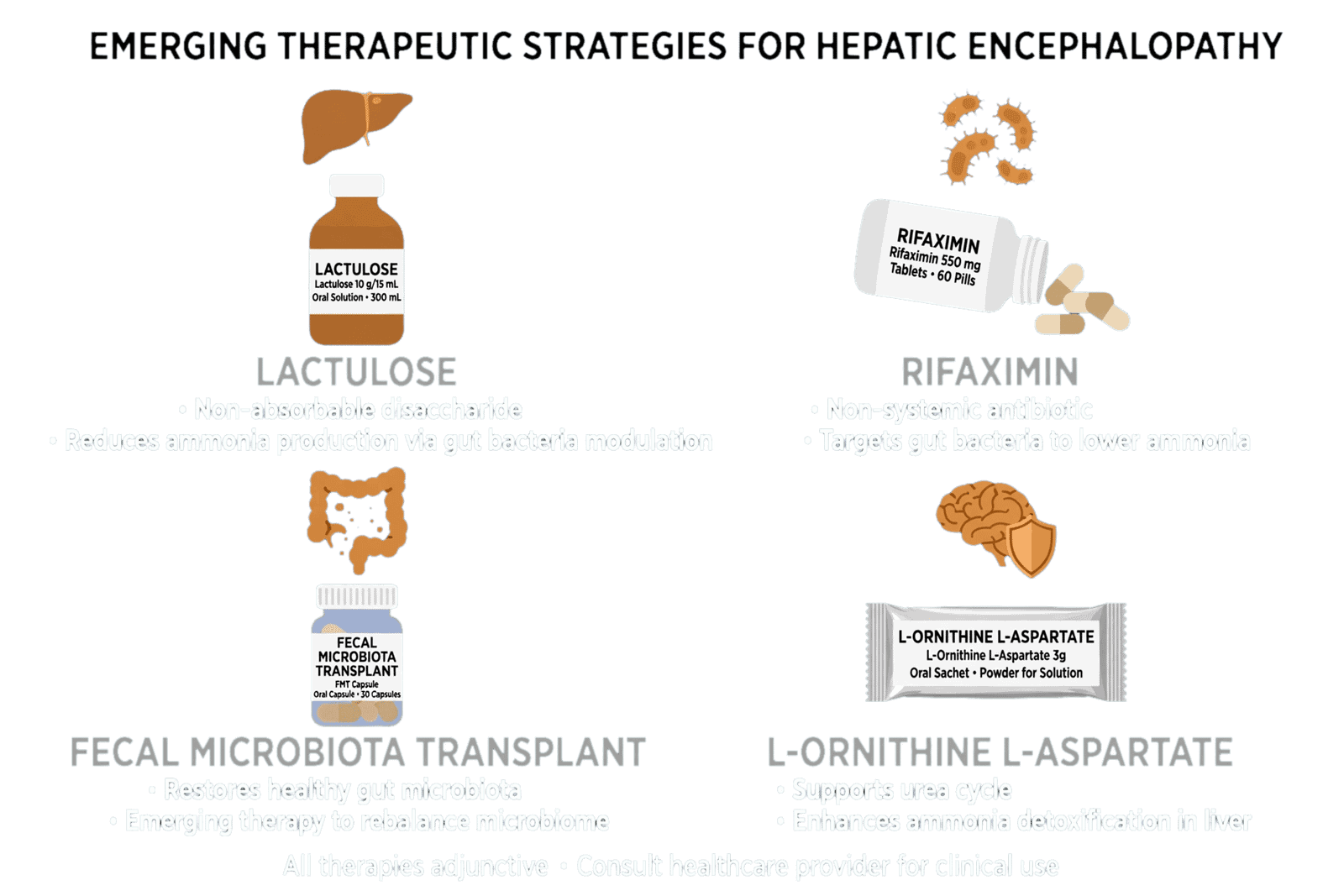
Other ammonia-lowering approaches include L-ornithine-L-aspartate and other nitrogen-handling strategies.
Evidence varies across therapies and regions, and not all treatments have the same level of guideline support.
Polyethylene glycol has generated particular interest for acute overt HE because rapid catharsis can produce faster improvement than conventional lactulose in selected hospitalized patients.
The HELP randomized trial compared PEG with lactulose in 50 hospitalized patients and found improvement of at least one HE grade at 24 hours in 91% of evaluable PEG-treated patients versus 52% receiving standard lactulose, with faster median resolution.
However, PEG should not automatically replace lactulose as universal standard therapy.
Trial size was limited, protocols varied, and patient selection matters.
More recent studies, including in acute-on-chronic liver failure, have also reported faster improvement with PEG-containing strategies, although broader implementation requires consideration of hydration, electrolyte status, aspiration risk, and local protocols.
The treatment of HE therefore increasingly resembles a layered strategy:
acute catharsis and ammonia reduction → correction of precipitating factors → maintenance therapy → nutritional and muscle preservation → prevention of recurrence.
Liver transplantation remains the definitive treatment for appropriate patients with advanced liver disease.
In patients with recurrent HE despite optimized medical therapy, transplant evaluation should not be delayed simply because the neurological symptoms are controlled temporarily.
Similarly, large spontaneous portosystemic shunts may require anatomical intervention in selected refractory cases.
The central limitation of established therapy is that much of it controls downstream manifestations rather than reversing the underlying cirrhosis.
This explains the continuing interest in microbiome therapeutics, ammonia scavengers, engineered bacteria, and other approaches.
The goal of emerging treatment is not simply to increase stool output.
It is to alter the biological ecosystem that generates and amplifies neurological dysfunction.
9. Emerging Therapeutic Strategies: Microbiome Engineering, FMT, Ammonia Scavengers, and Metabolic Therapies
The most exciting developments in hepatic encephalopathy are emerging from the recognition that the disorder involves multiple biological systems.
One of the most extensively studied approaches is fecal microbiota transplantation (FMT).
The rationale is straightforward: if dysbiosis contributes to HE, restoring a more favorable microbial ecosystem could potentially reduce neurotoxin production, improve intestinal barrier function, and modify systemic inflammation.
A landmark randomized clinical trial evaluated FMT from a carefully selected donor in men with cirrhosis and recurrent HE receiving standard therapy. The study found fewer serious adverse events, fewer further HE episodes, improved cognition, and favorable microbiome changes in the FMT group during follow-up.
These results were important but should not be interpreted as establishing FMT as routine standard therapy.
The trial was small.
Participants were selected.
Antibiotic pretreatment was used.
A specific donor was selected.
The intervention was performed under controlled conditions.
These characteristics make generalization difficult.
Safety also remains an important issue in cirrhosis because these patients may be particularly vulnerable to infection and bacterial translocation.
The future of microbiome therapy may therefore involve defined microbial consortia rather than conventional stool transplantation.
Engineered bacteria represent another intriguing possibility.
Instead of modifying the entire microbiome, researchers can potentially design microorganisms capable of consuming ammonia, altering nitrogen metabolism, producing beneficial metabolites, or suppressing specific harmful pathways.
This approach could offer greater reproducibility than FMT.
Another direction is targeted inhibition of microbial enzymes.
The recent discovery of phenylalanine decarboxylase activity associated with Ruminococcus gnavus and phenylethylamine production provides an example of how a specific microbial metabolic pathway could become a therapeutic target. Experimental targeting of this pathway reduced neurological abnormalities in models.
However, the transition from mechanistic discovery to human therapy is substantial.
Ammonia scavengers are another promising group.
Agents such as glycerol phenylbutyrate and ornithine phenylacetate attempt to increase nitrogen disposal through alternative metabolic pathways.
The principle is attractive because it targets systemic nitrogen handling rather than simply intestinal ammonia production.
Recent reviews identify nitrogen scavengers among the most important emerging pharmacological approaches under investigation.
L-ornithine-L-aspartate has also attracted considerable attention because it may enhance ammonia detoxification through urea-cycle and glutamine-synthesis pathways.
Albumin is another emerging adjunct.
Its potential benefits may extend beyond oncotic pressure and volume expansion, including modulation of oxidative stress, inflammatory signaling, and circulating toxins.
However, evidence is heterogeneous and albumin should not be viewed as a universal HE-specific therapy.
Other experimental approaches include agents targeting neurosteroid signaling, GABAergic pathways, oxidative stress, intestinal permeability, and ammonia metabolism.
GABA-A receptor-modulating steroid antagonists have been investigated because neurosteroid accumulation may contribute to altered inhibitory neurotransmission in cirrhosis.
Activated carbon-based therapies have also been explored as intestinal toxin-adsorbing strategies.
More recently, research has examined the possibility of replacing or enhancing ammonia-detoxifying enzymes such as glutamine synthetase.
These strategies are scientifically attractive because they address specific molecular defects.
Yet a critical principle must be maintained:
biological plausibility is not clinical efficacy.
A treatment that changes ammonia or microbiome composition does not necessarily improve cognition, reduce hospitalization, or improve survival.
Future trials should therefore prioritize clinically meaningful outcomes.
Cognitive function.
Overt HE recurrence.
Hospitalization.
Falls.
Quality of life.
Functional independence.
Transplant-free survival.
Mortality.
Biomarker improvement alone should not be sufficient.
The field also needs better patient selection.
A microbiome-targeted therapy may work best in patients whose HE phenotype is strongly driven by microbial dysfunction.
An ammonia scavenger may be more effective in patients with persistent nitrogen overload.
A portal-shunt intervention may be appropriate when anatomy is the dominant mechanism.
This is precision therapy.
Instead of asking:
“What is the best treatment for HE?”
the future question may become:
“What biological mechanism is dominant in this patient, and which intervention best targets it?”
That is a much more sophisticated therapeutic paradigm.
The current evidence supports optimism, but not premature adoption.
Lactulose and rifaximin remain the clinical foundation.
Emerging therapies should be viewed as complementary or investigational until supported by adequately powered, well-designed trials.
The ultimate goal is not to replace established therapy with fashionable technology.
It is to build a rational therapeutic hierarchy based on disease phenotype.
10. Precision Prevention and Personalized Long-Term Management
The most effective treatment for recurrent hepatic encephalopathy is often prevention.
An episode of HE should trigger a systematic evaluation of recurrence risk rather than simply a return to the previous treatment regimen.
The first layer is adherence.
Lactulose failure is frequently not pharmacological failure but practical failure.
Patients may stop therapy because of diarrhea, bloating, urgency, or social inconvenience.
Education must therefore explain the purpose of therapy and provide individualized titration instructions.
Rifaximin should be considered in appropriate patients with recurrent episodes despite standard therapy.
The landmark randomized trial demonstrated substantial reduction in recurrence with rifaximin, supporting its role in secondary prevention.
The second layer is precipitant prevention.
Patients and caregivers should understand early warning signs of infection, constipation, dehydration, gastrointestinal bleeding, medication changes, and altered mental status.
Caregivers are particularly important because patients may lose insight before they recognize cognitive deterioration themselves.
The third layer is nutrition and muscle preservation.
Adequate protein and energy intake should be maintained.
Prolonged fasting should be avoided.
Physical activity and resistance exercise may help preserve skeletal muscle when clinically appropriate.
This is not simply general wellness advice.
Muscle is part of ammonia metabolism.
Therefore, preserving muscle may be considered a component of ammonia management.
The fourth layer is medication optimization.
Benzodiazepines and unnecessary sedatives should be minimized.
Opioids should be carefully assessed.
Diuretics should be titrated to avoid excessive volume depletion.
Electrolytes and renal function should be monitored.
The fifth layer is anatomical assessment.
Patients with recurrent HE despite optimized medical therapy may require evaluation for large spontaneous portosystemic shunts or TIPS-related mechanisms.
In selected cases, embolization or revision may reduce recurrent HE.
The sixth layer is transplant assessment.
Recurrent HE is a major marker of decompensated cirrhosis and should prompt consideration of transplant eligibility.
The long-term goal is not merely fewer admissions.
It is restoration of functional independence and reduction of neurological burden.
This is particularly important for covert HE.
A patient who never develops overt coma may still experience major cognitive impairment.
Precision prevention therefore requires identifying these patients early.
Digital cognitive tools may eventually allow home monitoring.
A patient could perform a brief standardized cognitive task several times per week.
A progressive decline could trigger clinical review before overt HE develops.
Such systems would be particularly valuable for patients living far from specialist centers.
AI could potentially integrate cognitive trajectories with laboratory data, medication adherence, prior HE episodes, renal function, MELD-related variables, and microbiome or metabolomic information.
But again, implementation requires validation.
False alarms could create substantial burden.
Missed deterioration could create false reassurance.
Algorithms must therefore be prospectively tested in diverse populations.
Caregiver involvement should also be formalized.
Caregivers can recognize subtle changes in behavior, sleep, attention, and orientation.
They may be the first to identify recurrence.
Education should include a clear emergency plan.
The patient and family should know which symptoms require urgent hospital evaluation.
The long-term management model therefore becomes:
prevent → detect early → identify trigger → treat rapidly → restore baseline → reassess recurrence risk.
This is very different from the traditional model of waiting for overt HE and then administering lactulose.
The modern objective is to move intervention earlier in the disease trajectory.
Precision prevention may ultimately depend on identifying biological phenotypes.
For example:
A patient with recurrent infection-associated HE may benefit from aggressive infection surveillance.
A patient with profound sarcopenia may require intensive nutritional and physical rehabilitation.
A patient with large spontaneous shunting may need interventional assessment.
A patient with microbiome-associated metabolic abnormalities may eventually benefit from targeted microbial therapy.
A patient with repeated HE despite maximal medical treatment may require expedited transplant evaluation.
This approach avoids the assumption that every HE patient has the same disease mechanism.
The future of long-term HE care is therefore likely to be individualized.
The diagnosis will remain hepatic encephalopathy.
But the biological phenotype underneath that diagnosis may determine the optimal prevention strategy.
11. Future Directions: Biomarkers, Artificial Intelligence, Multi-Omics, and the Next Generation of HE Therapy
The next major transformation in hepatic encephalopathy is likely to come from the integration of clinical medicine with computational biology.
Current HE diagnosis depends heavily on clinical judgment because no single biomarker has adequate sensitivity, specificity, and reproducibility to replace bedside assessment.
This limitation creates an opportunity for multimodal precision diagnostics.
Future biomarker panels could integrate:
serum ammonia,
inflammatory cytokines,
microbial metabolites,
bile-acid profiles,
amino-acid signatures,
markers of astrocytic dysfunction,
renal function,
muscle mass,
and neurocognitive measurements.
Rather than asking whether one biomarker is elevated, clinicians could calculate an individualized probability of HE.
Metabolomics may be particularly useful.
The discovery of phenylethylamine as a potential microbial neurotoxin illustrates the concept that metabolites may sometimes be more biologically informative than microbial taxonomy alone.
A patient’s microbiome may contain thousands of microbial species.
But only a subset may be metabolically relevant to neurological disease.
Functional metabolomics could identify those pathways.
Artificial intelligence may then integrate these complex datasets.
An AI model could potentially recognize patterns associated with impending overt HE before symptoms become obvious.
It might combine:
recent hospitalization,
MELD-related variables,
ammonia trajectory,
renal function,
electrolytes,
lactulose adherence,
cognitive-test performance,
microbiome signatures,
and inflammatory markers.
The output could be a dynamic HE-risk score.
However, there are significant challenges.
Cirrhosis populations are heterogeneous.
Etiologies differ.
Treatment patterns differ.
Microbiomes differ geographically.
Socioeconomic factors affect adherence and hospitalization.
Therefore, an algorithm trained in one population may not generalize.
External validation must become a mandatory component of HE AI research.
Explainability is also important.
Clinicians need to know why an algorithm predicts high risk.
If the model identifies deteriorating renal function, increasing inflammation, and cognitive slowing as the dominant features, that information is clinically actionable.
If the model produces only an unexplained probability, implementation becomes more difficult.
Digital cognitive monitoring is another promising area.
Covert HE can be intermittent and difficult to detect during clinic visits.
Repeated home testing may reveal a patient’s personal cognitive baseline and detect meaningful deviation.
This could potentially reduce emergency presentations by allowing earlier intervention.
Wearable technologies may eventually contribute additional signals.
Changes in sleep architecture, activity, gait, reaction time, or autonomic function could potentially provide indirect indicators of neurological deterioration.
Again, these technologies require rigorous validation.
The future therapeutic landscape is equally dynamic.
Microbiome engineering could produce defined microbial consortia.
Engineered bacteria might consume ammonia or alter harmful metabolic pathways.
Targeted enzyme inhibition could block production of specific microbial neurotoxins.
Ammonia scavengers could enhance systemic nitrogen disposal.
Metabolic therapies could improve skeletal-muscle ammonia buffering.
Neuroprotective agents could reduce astrocyte vulnerability.
Combination therapies could target different levels of the disease simultaneously.
This is conceptually similar to oncology.
The future may not involve one universal HE treatment.
Instead, patients may receive mechanism-specific combinations.
The research field should therefore move toward phenotype-based clinical trials.
A trial of a microbiome therapy should identify patients with microbiome dysfunction.
A trial of an ammonia scavenger should characterize nitrogen metabolism.
A trial of a neuroinflammatory therapy should identify patients with relevant inflammatory signatures.
This would improve the probability of detecting clinically meaningful treatment effects.
The emerging gut–liver–brain paradigm also creates opportunities for preventive medicine.
If a patient is identified as high-risk before overt HE, intervention may become more effective.
This is particularly important because recurrent HE can produce cumulative cognitive and functional consequences.
Ultimately, the most transformative development may not be a new drug.
It may be the ability to identify the dominant biological mechanism in each patient and intervene before the clinical syndrome becomes severe.
The recent expansion of HE research into microbiome-derived neurotoxins, multi-organ interactions, and precision diagnostics suggests that the field is moving in this direction.
The next generation of HE medicine will therefore likely be defined by integration.
Clinical hepatology.
Neuroscience.
Microbiology.
Metabolomics.
Nutrition.
Digital medicine.
Artificial intelligence.
Transplantation.
The objective is not to abandon the ammonia hypothesis.
It is to finally understand what happens around it.
12. Conclusions: From an Ammonia-Centered Disease Model to Systems-Level Precision Hepatology
Hepatic encephalopathy is no longer adequately described as a neurological consequence of elevated ammonia alone.
Ammonia remains a major neurotoxin and an essential component of HE pathophysiology, but contemporary evidence demonstrates that neurological dysfunction emerges from a complex interaction among hyperammonemia, portal-systemic shunting, systemic inflammation, intestinal dysbiosis, microbial metabolites, impaired intestinal barrier function, sarcopenia, renal dysfunction, electrolyte abnormalities, astrocyte dysfunction, and cerebral vulnerability.
The gut–liver–brain axis provides a more complete framework for understanding this complexity.
The intestine generates ammonia and numerous other metabolites.
The diseased liver fails to detoxify them adequately.
Portal-systemic shunts bypass hepatic metabolism.
The immune system responds to microbial translocation.
Skeletal muscle loses its capacity to buffer nitrogen.
The brain becomes increasingly vulnerable to metabolic and inflammatory stress.
The clinical manifestation is hepatic encephalopathy.
This framework also explains why serum ammonia is insufficient as a stand-alone diagnostic test.
The AASLD emphasizes that ammonia concentrations do not reliably correlate with HE grade and that HE remains a clinical diagnosis.
Precision diagnosis therefore requires clinical assessment, appropriate cognitive testing, exclusion of alternative causes of altered mental status, and selective use of biomarkers and neurodiagnostic tools.
Covert HE deserves particular attention because subtle cognitive dysfunction can significantly affect safety, quality of life, independence, and progression toward overt disease.
Established therapy remains centered on lactulose and rifaximin, supported by correction of precipitating factors and appropriate nutritional management.
Rifaximin has strong randomized-trial evidence for reducing recurrent HE.
Rapid gut catharsis with PEG may accelerate recovery in selected acute presentations, although it should complement rather than automatically replace individualized standard therapy.
The emerging therapeutic landscape is considerably broader.
FMT, engineered bacteria, microbial enzyme inhibition, ammonia scavengers, metabolic therapies, albumin-based approaches, and neuroactive pathway modulation are all being investigated.
The early FMT randomized trial demonstrated encouraging improvements in recurrent HE, cognition, and microbiome characteristics, but its limited population and specialized protocol emphasize why larger trials remain necessary before routine widespread adoption.
Perhaps the most important future development is the move toward biological phenotyping.
Not all HE is biologically identical.
One patient may be dominated by ammonia accumulation.
Another by systemic inflammation.
Another by sarcopenia.
Another by large portal-systemic shunting.
Another by microbial neurotoxins.
Another by several mechanisms simultaneously.
Precision hepatology seeks to identify these differences and use them therapeutically.
The ultimate transition is therefore from:
“Does this patient have hepatic encephalopathy?”
toward:
“Which biological mechanisms are driving this patient’s encephalopathy, and which interventions can modify those mechanisms?”
That is the central promise of the gut–liver–brain paradigm.
HE is increasingly becoming a model disease for systems medicine: a condition in which the liver, intestine, immune system, skeletal muscle, circulation, and brain function as an interconnected biological network.
The future will not be defined by abandoning ammonia.
It will be defined by understanding what ammonia does, why the brain becomes vulnerable to it, how the gut modifies its effects, and how the entire system can be therapeutically redirected.
The most competitive future research in HE will therefore lie at the intersection of microbiome science, neurobiology, metabolomics, precision diagnostics, artificial intelligence, and personalized therapeutics.
The disease is no longer simply an ammonia problem.
It is a systems problem.
And the therapeutic opportunity lies precisely in understanding the system.