




Introduction: From Conventional Ablation to Precision Electrophysiology
Cardiac electrophysiology has undergone a fundamental transformation over the past several decades. Early catheter ablation strategies were largely guided by anatomical landmarks, conventional intracardiac electrograms, and relatively limited spatial sampling. As three-dimensional electroanatomical mapping systems developed, electrophysiologists gained the ability to reconstruct cardiac chambers, visualize catheter position, create activation maps, identify low-voltage regions, and reduce dependence on fluoroscopy. These developments substantially improved procedural precision. Yet contemporary arrhythmia treatment still frequently relies on simplified disease categories such as paroxysmal versus persistent atrial fibrillation, typical versus atypical atrial flutter, or ischemic versus nonischemic ventricular tachycardia. Such classifications are clinically useful, but they do not fully capture the biological heterogeneity of individual arrhythmogenic substrates.
Precision electrophysiology represents an emerging attempt to address this limitation by integrating multiple layers of patient-specific information. Instead of considering electrophysiological mapping, imaging, artificial intelligence, and ablation energy as independent technologies, precision electrophysiology treats them as components of a continuous decision-making system. Anatomical imaging describes the patient’s cardiac architecture. Structural imaging can identify fibrosis, scar, chamber remodeling, and tissue heterogeneity. High-density mapping provides electrical information at substantially greater spatial and temporal resolution. Artificial intelligence can process large multidimensional datasets and potentially identify patterns that are difficult to recognize through conventional visual interpretation. Finally, advanced ablation technologies such as pulsed-field ablation can translate a selected treatment strategy into rapid, controlled tissue modification.
The concept is particularly relevant to atrial fibrillation. The pulmonary veins remain fundamental targets, but AF is not a uniform electrical disorder. Patients may have different combinations of pulmonary venous triggers, atrial fibrosis, conduction slowing, reentry, non-pulmonary-vein triggers, autonomic influences, and structural remodeling. Two patients with identical clinical labels may therefore have very different electrophysiological substrates. The 2024 international AF ablation consensus recognizes the growing role of electroanatomical mapping, preprocedural imaging, MRI-based fibrosis characterization, ECG imaging, and emerging computational approaches while also emphasizing unresolved questions regarding the clinical value of some advanced mapping strategies. [1]
High-density mapping has already increased the amount of electrical information available during procedures. Contemporary multipolar catheters can acquire thousands of points efficiently, allowing more detailed visualization of voltage and activation patterns. An observational Asia-Pacific study involving 200 AF patients demonstrated the feasibility of efficient high-density mapping, with a median mapping time of approximately nine minutes. [2] The technological objective is not simply to collect more points but to reduce sampling error and better characterize spatially heterogeneous electrical activity.
Cardiac imaging adds another dimension. CT can provide high-resolution anatomical information, pulmonary vein anatomy, atrial geometry, and procedural planning data. MRI provides soft-tissue characterization and can identify atrial fibrosis through late gadolinium enhancement. The relationship between atrial fibrosis and AF recurrence has been demonstrated in multiple studies, although the clinical utility of fibrosis-guided ablation remains more controversial than fibrosis-based risk stratification. DECAAF demonstrated an association between increasing fibrosis burden and recurrence, whereas DECAAF II did not establish that routine MRI-guided fibrosis ablation improved outcomes when added to PVI. [3–5]
Artificial intelligence introduces a further layer. Machine-learning systems can integrate clinical variables, imaging features, ECG characteristics, and electrophysiological measurements to estimate recurrence risk or identify phenotypic subgroups. However, AI performance is highly dependent on training populations, endpoint definitions, data quality, and external validation. A recent study combining CT-derived features and clinical variables achieved an AUC of 0.76 for predicting one-year AF recurrence, illustrating potential but also demonstrating that prediction is far from perfect. [6]
Pulsed-field ablation completes the conceptual chain by providing a nonthermal energy platform that can rapidly modify myocardium. Its major clinical attraction is not simply speed but a distinct tissue-injury mechanism and favorable profile for several collateral structures.
The ultimate objective of precision electrophysiology is therefore not technological accumulation. It is the creation of a patient-specific workflow in which each dataset answers a different question: Where is the substrate? What is it doing electrically? Which components are clinically relevant? Which targets should be treated? How should energy be delivered? And did the intervention actually produce the intended biological effect?
This transition—from mapping what is visible to understanding what is biologically relevant—may define the next major phase of cardiac electrophysiology.
The Biological and Clinical Rationale for Personalized Arrhythmia Treatment
The fundamental rationale for precision electrophysiology is that arrhythmias are phenotypically diverse diseases rather than single uniform entities. Atrial fibrillation provides the clearest example. Although pulmonary venous ectopy is a central mechanism in many patients, AF persistence can involve atrial fibrosis, chamber enlargement, altered conduction, reentrant activity, autonomic remodeling, inflammation, metabolic disease, and interactions with systemic cardiovascular risk factors.
A conventional population-based approach assigns patients to broad categories and applies a relatively standardized lesion set. This strategy has substantial value because pulmonary vein isolation is reproducible, evidence-based, and effective. However, the remaining recurrence after ablation demonstrates that anatomical classification alone cannot explain every patient’s disease behavior.
Personalization begins with recognizing heterogeneity. A patient with small atria, limited fibrosis, short AF duration, and predominantly pulmonary venous triggers is biologically different from a patient with extensive atrial remodeling, diffuse fibrosis, persistent AF, enlarged atria, and multiple non-pulmonary-vein drivers. Both may carry the same diagnostic label, but their probability of durable rhythm control and their optimal procedural strategy may differ.
Cardiac MRI provides an example of this biological stratification. The original DECAAF study demonstrated a graded relationship between left atrial fibrosis burden and arrhythmia recurrence after ablation. Patients with more advanced fibrosis had substantially higher recurrence rates. [3] Five-year follow-up similarly demonstrated an association between advanced fibrosis and recurrent arrhythmia and repeat ablation. [4]
However, the failure of fibrosis-guided ablation in DECAAF II to demonstrate improved outcomes compared with PVI alone is equally important. [5] This finding illustrates a critical principle of precision medicine: identifying a biomarker associated with prognosis does not automatically mean that treating the biomarker improves outcomes. A predictive marker can identify risk without identifying a therapeutically actionable target.
This distinction applies equally to electrophysiological mapping. Low-voltage areas, complex fractionated electrograms, rotational activity, focal impulses, conduction slowing, and dispersion patterns may all represent biologically interesting phenomena. But the existence of an abnormal electrical signal does not prove that eliminating that signal improves clinical outcomes.
Precision electrophysiology therefore requires three separate concepts: characterization, prediction, and intervention. Characterization describes the patient’s substrate. Prediction estimates what may happen. Intervention attempts to change the trajectory. Confusing these three stages is a major source of overinterpretation.
The biological rationale also extends beyond AF. Ventricular tachycardia after myocardial infarction may involve dense scar, border zones, channels of surviving myocardium, and heterogeneous conduction. Nonischemic cardiomyopathy can involve patchy fibrosis that is difficult to characterize by conventional voltage mapping alone. Congenital heart disease can produce highly complex anatomical and surgical substrates. In each scenario, combining anatomical imaging with electrical mapping may provide a more complete representation than either modality independently.
Personalized treatment also requires attention to the patient’s clinical context. Obesity, hypertension, diabetes, sleep apnea, alcohol exposure, physical inactivity, and structural heart disease influence atrial remodeling and recurrence. The 2024 consensus emphasizes risk-factor modification as an important component of AF ablation management. [1]
This means that precision electrophysiology should not be reduced to a sophisticated catheter procedure. It should encompass preprocedural phenotyping, procedural targeting, postprocedural monitoring, and long-term modification of the biological environment that promotes arrhythmia.
The ultimate objective is not maximum information. It is actionable information.
An ideal precision workflow would therefore classify each patient according to the dominant mechanism, quantify structural disease, characterize electrical behavior, estimate recurrence risk, select the minimum effective lesion set, choose an energy source appropriate to the anatomy, confirm lesion success, and continuously reassess the patient’s rhythm after intervention.
Such a strategy could theoretically reduce unnecessary ablation while directing additional treatment toward patients with biologically meaningful substrate. But this remains a hypothesis for many advanced strategies. The field must continue to distinguish between technologies that produce visually impressive maps and technologies that demonstrably improve patient outcomes.
Precision electrophysiology becomes clinically meaningful only when increased resolution changes management in a way that improves outcomes, safety, or efficiency.
High-Density Electroanatomical Mapping: Increasing the Resolution of the Arrhythmogenic Substrate
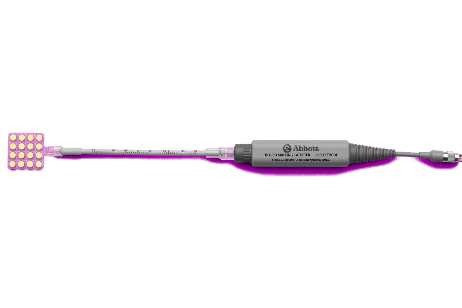
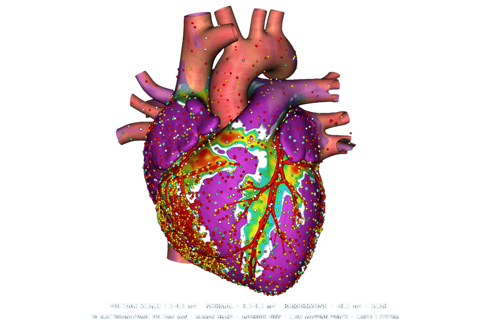
High-density electroanatomical mapping represents one of the most important technological foundations of precision electrophysiology. Traditional mapping samples electrical activity from a relatively limited number of locations. Although interpolation can generate visually continuous maps, sparse acquisition may miss narrow conduction channels, localized scar, small reentrant circuits, or rapidly changing activation patterns. High-density mapping attempts to overcome this limitation by simultaneously acquiring signals from multiple electrodes distributed across flexible multipolar catheters.
The conceptual advantage is straightforward: more spatial samples should allow a more faithful representation of cardiac electrophysiology. In practice, however, the value of high-density mapping depends on signal quality, catheter stability, electrode spacing, filtering, annotation algorithms, and the physiological state during acquisition.
Modern high-density systems can generate detailed voltage maps that identify normal myocardium, low-voltage regions, and electrical scar. They can also construct activation maps that reveal conduction direction, activation timing, block lines, and potential reentrant pathways. In complex atrial tachycardias, these capabilities can be particularly valuable because a narrow isthmus may represent the critical component sustaining an otherwise extensive circuit.
High-density mapping also improves the ability to revisit previously treated regions. Patients undergoing redo procedures may have patchy reconnection around pulmonary veins or organized atrial tachycardias arising from prior lesion sets. A dense map can identify conduction gaps more precisely than conventional sampling.
A prospective observational study of 200 patients across the Asia-Pacific region demonstrated the feasibility of high-density mapping in AF procedures, with a median mapping time of approximately nine minutes. The study also showed that pulmonary vein isolation remained universal while non-pulmonary-vein targets were selected in a subset of patients. [2]
Yet high-density mapping creates a paradox: more data do not necessarily produce better decisions.
The first problem is data interpretation. A map containing tens of thousands of points can reveal subtle abnormalities, but some may represent noise, far-field signals, catheter instability, poor contact, or differences in rhythm state rather than genuine substrate. High-density mapping therefore increases the need for sophisticated quality control.
The second problem is temporal variability. AF is not electrically static. Activation patterns may change from beat to beat. A region appearing as a driver at one moment may not behave similarly several seconds later. This raises questions about whether a high-resolution map represents a stable biological substrate or merely a transient electrical configuration.
The third problem is clinical relevance. The 2024 international consensus discusses technologies capable of identifying complex activation, charge-density patterns, and spatiotemporal dispersion, but explicitly highlights unresolved questions regarding whether targeting these findings improves outcomes. [1]
This is especially important in persistent AF. The ability to identify focal impulses, rotational patterns, or dispersed electrograms is scientifically attractive. However, targeting every abnormal region risks creating excessive scar without clear evidence of benefit.
High-density mapping is therefore most powerful when used to answer a specific question. In atrial tachycardia, it can identify the circuit and critical isthmus. In redo AF procedures, it can identify reconnection. In ventricular tachycardia, it can delineate scar channels and late potentials. During PVI, it can provide high-resolution confirmation of electrical isolation.
Another major development is integration with imaging. Electroanatomical maps can be registered with CT or MRI-derived anatomy, allowing electrical abnormalities to be interpreted within a structural context. A low-voltage region located within an MRI-defined fibrotic zone may have different significance from an isolated low-voltage point without structural correlate.
Future mapping systems will likely incorporate increasingly automated signal classification. Artificial intelligence could assist in identifying abnormal electrograms, conduction abnormalities, or potential drivers. The goal should not be to replace the electrophysiologist but to reduce the cognitive burden associated with interpreting massive datasets.
High-density mapping is therefore best understood as an information amplifier. It increases spatial resolution, but the clinical value comes from translating that information into reproducible biological interpretation.
The next evolution will not simply be “higher density.” It will be higher-confidence density: maps in which every clinically relevant point has reliable signal quality, anatomical registration, temporal context, and a defined relationship to the arrhythmia mechanism.
That distinction will determine whether high-density mapping becomes merely a technological visualization tool or a genuine foundation of precision electrophysiology.
Cardiac Imaging as a Structural and Anatomical Layer of Electrophysiological Mapping
Electrophysiology traditionally describes electrical behavior, whereas cardiac imaging describes anatomy and tissue structure. Precision electrophysiology requires both because electrical abnormalities cannot always be interpreted correctly without understanding the substrate in which they occur.
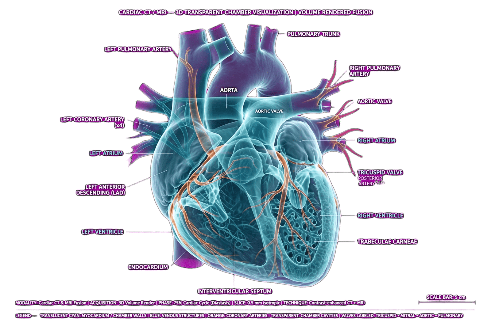
Cardiac computed tomography provides high-resolution anatomical information and is particularly useful for pulmonary vein anatomy, left atrial geometry, chamber volume, and procedural planning. CT can be integrated with three-dimensional electroanatomical maps, allowing the operator to navigate according to patient-specific anatomy rather than relying exclusively on generic chamber geometry.
This becomes particularly relevant when pulmonary vein anatomy is complex. Common variations include accessory veins, common ostia, early branching, and unusual orientations. Understanding this anatomy before catheter manipulation can improve procedural planning and reduce unnecessary energy delivery.
Magnetic resonance imaging provides a different advantage: tissue characterization. Late gadolinium enhancement can identify areas of atrial fibrosis and ventricular scar. In AF, left atrial fibrosis is associated with disease progression and recurrence after ablation. The DECAAF studies demonstrated that fibrosis burden provides prognostic information, although the therapeutic strategy of directly ablating MRI-defined fibrosis has not been conclusively validated. [3–5]
This distinction is central to precision medicine. Imaging can tell us that a patient has a high-risk substrate without necessarily telling us which tissue should be ablated.
MRI can also contribute to ventricular arrhythmia management. In ischemic cardiomyopathy, infarct scar can be delineated anatomically, while in nonischemic cardiomyopathy, fibrosis distribution may reveal potential arrhythmogenic substrate. Integration of MRI-derived scar with intracardiac voltage mapping can therefore create a multimodal representation of the substrate.
Electrocardiographic imaging adds another dimension. Unlike intracardiac mapping, ECG imaging can provide noninvasive information about electrical activation across large regions of the atria and ventricles. The 2024 consensus describes ECGI as a noninvasive approach capable of simultaneous global characterization of biatrial electrical activity, although its spatial resolution differs from invasive mapping. [1]
The value of imaging therefore lies in complementarity rather than replacement.
CT answers: What does the anatomy look like?
MRI asks: What is the tissue composition?
Electrophysiological mapping asks: How does the tissue conduct?
ECGI asks: How does electrical activation behave across the organ?
AI may ultimately ask: How do these variables interact to predict this patient’s disease trajectory?
The integration of these modalities is technically challenging. Registration errors can arise because CT or MRI is acquired at a different respiratory phase, volume status, rhythm, or cardiac cycle than the invasive procedure. The heart moves, the atria deform, and catheter-induced changes can alter local anatomy. The 2024 consensus specifically recognizes multimodality integration but also notes the practical limitations of registration accuracy. [1]
Imaging quality is another limitation. LGE-MRI requires specialized acquisition protocols and expertise, and poor image quality can prevent reliable fibrosis quantification. The original DECAAF study excluded a meaningful proportion of patients because of inadequate MRI quality. [3]
Radiation is another consideration for CT, although modern protocols can substantially reduce exposure. MRI avoids ionizing radiation but can require longer acquisition times and may not be suitable for all patients.
The future of precision electrophysiology will probably involve computational segmentation of these images. AI algorithms can automatically identify atrial boundaries, pulmonary veins, scar, fibrosis, ventricular geometry, and other structures. These datasets can then be transferred into electroanatomical mapping systems.
The ultimate goal is a patient-specific digital anatomical model that exists before the procedure begins.
However, imaging should not be treated as an oracle. A beautiful MRI fibrosis map can be wrong, an anatomical registration can be imperfect, and an imaging biomarker can be prognostic without being therapeutically actionable.
Therefore, imaging should serve as a structural layer within a multimodal model rather than dictating ablation independently.
Its greatest potential may be realized when anatomical, structural, and electrical abnormalities converge. When imaging identifies fibrosis, high-density mapping demonstrates conduction abnormalities in the same region, and the patient’s clinical phenotype supports that substrate as mechanistically relevant, the combined evidence becomes more compelling than any single modality alone.
Artificial Intelligence and Machine Learning in Modern Cardiac Electrophysiology
Artificial intelligence is increasingly entering cardiac electrophysiology because arrhythmia care generates unusually large and heterogeneous datasets. A single patient may produce ECG recordings, ambulatory rhythm data, echocardiography, CT, MRI, intracardiac electrograms, three-dimensional maps, procedural data, laboratory values, medication histories, and long-term follow-up information. Human interpretation of all these variables simultaneously is difficult.
AI offers a potential method for integrating these data.
Machine-learning systems can identify nonlinear relationships between variables that may be difficult to recognize through conventional statistical modeling. Deep-learning models can analyze raw ECG signals or medical images. Natural-language and multimodal systems may eventually integrate clinical records with structured physiological information.
One important application is preprocedural risk prediction. A 2024 study developed AI models combining pulmonary-vein CT images with clinical variables to predict one-year AF recurrence after ablation. The CT-only model achieved an AUC of 0.63, while an ensemble combining imaging and clinical information reached an AUC of 0.76. [6] This illustrates both the potential and the limitation of AI: combining complementary information can improve prediction, but prediction remains probabilistic rather than deterministic.
Another application is electrophysiological signal interpretation. High-density mapping produces enormous quantities of electrograms. AI algorithms can potentially identify fractionation, conduction slowing, dispersion, focal activity, or other patterns automatically. The 2024 consensus discusses machine-learning approaches for identifying spatiotemporal dispersion and other potential AF targets, while noting that stability and reproducibility of these targets require further validation. [1]
AI can also support treatment stratification. Recent work using ECGI-derived electrophysiological complexity with clinical information has shown that machine-learning clustering can identify groups with different outcomes after rhythm-control strategies. [7]
However, AI should not be equated with accuracy.
A model may perform exceptionally well in its development population but poorly in another hospital because of differences in patient demographics, equipment, acquisition protocols, disease prevalence, or treatment strategy. This phenomenon, known as dataset shift, is particularly important in medical AI.
Explainability is another challenge. An electrophysiologist may reasonably ask why an algorithm identified a particular region as a potential ablation target. If the answer cannot be meaningfully explained, clinical adoption may be limited.
The problem is especially important when AI influences irreversible interventions. A recommendation to prescribe a medication can be revisited. A recommendation to ablate tissue creates permanent biological change.
Therefore, AI-supported ablation should initially function as decision support rather than autonomous decision-making.
Another major issue is endpoint selection. An AI system trained to predict recurrence may optimize statistical prediction without identifying a modifiable mechanism. A patient may be correctly classified as high risk while the model provides no actionable target.
This distinction separates prognostic AI from therapeutic AI.
The ideal precision-electrophysiology AI system would not simply predict that recurrence is likely. It would explain which biological factors contribute to the risk, identify potentially modifiable mechanisms, estimate the expected benefit of alternative lesion strategies, and provide uncertainty estimates.
Future systems may therefore become multimodal. Rather than analyzing CT, MRI, ECG, and electrograms separately, an integrated model could process all modalities simultaneously. This could produce a patient-specific electrophysiological phenotype.
Digital twins represent the most ambitious version of this concept. A digital twin would be a computational representation of the patient’s heart capable of simulating activation, conduction, scar, and potentially the predicted effects of different ablation strategies.
This remains an emerging research direction rather than routine clinical practice.
The appropriate current position is therefore balanced. AI has genuine potential to improve electrophysiological diagnosis, mapping interpretation, risk prediction, and procedural planning. But the field still requires external validation, prospective trials, transparency, reproducibility, and evidence that algorithmic assistance improves patient outcomes rather than merely improving retrospective metrics.
Precision electrophysiology should use AI to manage complexity—not to create a new layer of unexplained complexity.
Pulsed-Field Ablation: Translating Precision Mapping Into Tissue-Selective Therapy
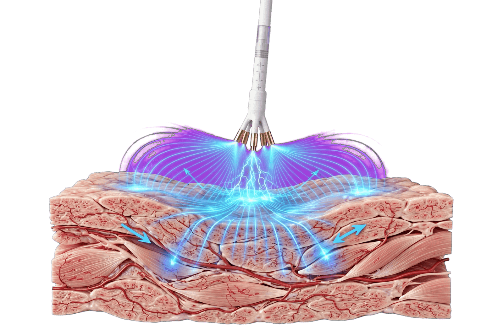
Pulsed-field ablation represents a major change in how electrophysiological information can be translated into tissue treatment. Conventional radiofrequency ablation relies on thermal energy, whereas cryoablation relies on freezing. PFA uses short, high-voltage electrical pulses to induce irreversible electroporation and cellular injury.
The significance of PFA within precision electrophysiology is its potential combination of rapid treatment, controllable lesion geometry, and relative tissue selectivity.
This becomes particularly important after high-resolution mapping has identified the intended target. Precision treatment requires not only identifying abnormal tissue but also selecting an energy source that can modify that tissue while minimizing injury to adjacent structures.
Clinical evidence has established PFA as an effective technology for AF ablation. The ADVENT randomized trial demonstrated noninferiority of PFA compared with conventional thermal ablation in paroxysmal AF. Large real-world datasets have subsequently demonstrated high procedural success and low rates of several classic thermal complications.
The 2024 international consensus recognizes PFA as a major new ablation modality and describes advantages including procedural efficiency and a favorable safety profile for certain collateral structures. [1]
The biological concept is particularly relevant to precision therapy. Electroporation is influenced by electric-field strength, pulse duration, waveform, electrode configuration, tissue characteristics, and catheter-tissue interaction. Therefore, PFA is not simply an energy source that is delivered identically everywhere. Its effect depends on how the field interacts with anatomy.
This creates a natural relationship between mapping and PFA.
High-density mapping identifies where electrical isolation or substrate modification is required.
Imaging identifies surrounding structures and tissue architecture.
AI can potentially help determine which targets are most clinically relevant.
PFA then provides a rapid mechanism for executing the selected treatment.
The safety profile is also important. Large registry data have demonstrated very low observed rates of esophageal injury, pulmonary vein stenosis, and persistent phrenic nerve injury. However, PFA does not eliminate complications. Coronary spasm and hemolysis have emerged as energy-specific concerns, while tamponade, vascular injury, and thromboembolic complications remain possible because the overall procedure still requires intracardiac instrumentation. [8]
Therefore, precision PFA should not mean “more ablation because PFA is safer.” That would be a dangerous interpretation.
Instead, improved mapping should allow the operator to identify the minimum lesion set necessary for the intended therapeutic objective.
This is particularly relevant in persistent AF. The ease with which PFA can treat large areas makes posterior wall isolation technically attractive. Yet technical feasibility does not establish clinical necessity. A large lesion set may create unnecessary scar if the additional target does not contribute meaningfully to the patient’s arrhythmia mechanism.
Precision therefore requires restraint.
Another important issue is lesion assessment. PFA can produce acute electrogram disappearance, but immediate electrical silence may include reversible stunning. Confirmation of durable isolation remains essential.
PFA also offers opportunities for complex arrhythmias. Studies have explored its use for post-ablation atrial tachycardias and other substrates. One comparative study of post-AF-ablation left atrial tachycardias found PFA to be faster than RF but less effective at one-year follow-up in that particular cohort, illustrating that the performance of PFA may depend heavily on the arrhythmia substrate and catheter design. [9]
This is precisely why precision electrophysiology must integrate energy selection with substrate characterization.
PFA is not automatically superior for every arrhythmia.
Its strongest current role is in AF, particularly pulmonary vein isolation, where its combination of efficiency and safety has substantial clinical support. Broader applications remain under investigation.
The future may involve adaptive PFA in which mapping systems determine the exact electrical field and application pattern required for each anatomical region. Instead of delivering a standardized number of applications, the system could theoretically respond to tissue-specific feedback.
Such a system would represent a true convergence of mapping, imaging, AI, and energy delivery.
PFA therefore should be viewed not as the endpoint of precision electrophysiology but as its current therapeutic engine: the mechanism by which increasingly sophisticated patient-specific information can ultimately be translated into controlled biological intervention.
Multimodal Data Fusion: Building a Patient-Specific Electrophysiological Model
The central challenge of precision electrophysiology is not obtaining individual technologies. Modern EP laboratories may already possess high-density mapping, CT integration, MRI, intracardiac echocardiography, automated annotation, and advanced ablation systems. The challenge is making these technologies communicate with one another.
Multimodal data fusion attempts to solve this problem.
A patient-specific model could theoretically contain four major layers.
The first is anatomical. CT or MRI defines the chamber geometry, pulmonary veins, valves, appendages, ventricular structures, and other relevant anatomy.
The second is structural. MRI can characterize fibrosis or scar, while echocardiography can quantify chamber size, function, strain, and hemodynamic features.
The third is electrical. High-density mapping identifies voltage, activation timing, conduction pathways, electrical scar, and potential arrhythmogenic regions.
The fourth is clinical and temporal. AF duration, comorbidities, ECG characteristics, medication response, rhythm burden, previous procedures, and longitudinal outcomes provide context.
AI can potentially integrate these layers.
Consider a patient with persistent AF. CT identifies a markedly enlarged left atrium. MRI demonstrates substantial fibrosis. High-density mapping identifies multiple low-voltage regions and conduction heterogeneity. ECGI suggests increased electrical complexity. Clinical history reveals long-standing AF and multiple cardiovascular risk factors.
Individually, none of these findings provides a complete treatment plan.
Together, they define a phenotype of advanced atrial remodeling.
The opposite phenotype might include a relatively small atrium, limited fibrosis, short-duration AF, preserved ventricular function, and predominantly pulmonary venous triggers. Such patients may require a very different procedural strategy.
This illustrates the central principle of multimodal precision: the patient is the unit of analysis, not the technology.
However, multimodal integration introduces new technical problems.
The first is registration. Imaging acquired before the procedure must be aligned with anatomy reconstructed during the procedure. Respiratory motion, cardiac motion, volume changes, and differences in rhythm can create spatial discrepancies.
The second is scale. MRI fibrosis may be represented as tissue-level information, whereas intracardiac mapping samples localized electrical signals. These datasets operate at different spatial resolutions.
The third is temporal mismatch. Imaging may represent anatomy from days or weeks before ablation, whereas electrophysiology reflects the patient’s real-time electrical state.
The fourth is uncertainty. Every modality contains measurement error. Combining multiple imperfect datasets can improve confidence, but it can also propagate errors.
Therefore, multimodal systems require uncertainty-aware integration rather than simply overlaying images.
A clinically useful system should ideally distinguish high-confidence findings from uncertain predictions. For example, a pulmonary vein ostium identified clearly on CT represents a different level of certainty from an AI-generated prediction that a particular region is an AF driver.
This distinction becomes particularly important when irreversible ablation is planned.
One potential solution is probabilistic modeling. Instead of defining a single “target,” the system could generate a map of target probability based on structural, electrical, and clinical evidence.
Another possibility is dynamic mapping. Rather than using a static preprocedural model, real-time electrograms and intracardiac imaging could continuously update the patient-specific representation.
This could eventually create a procedural “digital twin.”
The digital twin would not need to reproduce every biological process. Its clinical value would come from accurately representing the variables relevant to ablation decisions.
For example, it could simulate the likely electrical effect of isolating a pulmonary vein, modifying a conduction channel, or treating a posterior wall region.
Such systems remain experimental, but the conceptual direction is important.
The goal of multimodal fusion is not to produce the most complicated map.
It is to reduce uncertainty.
If imaging reduces anatomical uncertainty, high-density mapping reduces electrical uncertainty, AI reduces interpretive uncertainty, and PFA reduces uncertainty about collateral thermal injury, the combined system may allow more targeted intervention.
This represents the true promise of precision electrophysiology.
The future EP laboratory may therefore resemble a data-integration environment as much as a traditional catheter laboratory. The operator will not simply manipulate a catheter while interpreting electrograms. Instead, the operator will interact with a continuously updated patient-specific model that combines anatomy, tissue, electrical activity, prediction, and treatment response.
That transformation will only be clinically meaningful if it ultimately improves outcomes, reduces unnecessary ablation, decreases complications, or makes complex procedures more reproducible.
Personalized Ablation Strategies for Atrial Fibrillation and Complex Atrial Arrhythmias
Atrial fibrillation is currently the most mature clinical setting for precision electrophysiology because it combines heterogeneous substrate, large patient populations, established ablation endpoints, and rapidly developing technologies.
Pulmonary vein isolation remains the foundation of AF ablation. The reason is not technological conservatism but strong biological and clinical evidence demonstrating the importance of pulmonary venous triggers.
Precision electrophysiology does not therefore mean abandoning PVI.
Instead, it asks whether the patient’s characteristics justify anything beyond PVI and whether additional intervention can be targeted more intelligently.
In paroxysmal AF with limited structural remodeling, the personalized strategy may be relatively focused. High-density mapping can confirm pulmonary vein anatomy and isolation. CT can define anatomical variants. Intracardiac echocardiography can assist procedural guidance. PFA can provide rapid PVI while reducing several thermal collateral risks.
In persistent AF, the problem becomes more complex. The atrial substrate may include fibrosis, low-voltage areas, conduction slowing, non-PV triggers, and organized reentrant activity.
MRI may provide prognostic information about fibrosis. High-density mapping can characterize electrical heterogeneity. AI may potentially classify patients according to recurrence risk or electrophysiological complexity. The challenge is translating these findings into an evidence-based lesion set.
This is where precision electrophysiology must resist the temptation of technological determinism.
If a map shows a low-voltage region, the appropriate response is not automatically to ablate it.
If MRI shows fibrosis, the appropriate response is not automatically to destroy it.
If AI identifies a potential driver, the appropriate response is not automatically to deliver energy there.
Each proposed target must pass a causal test: Is the abnormality mechanistically related to the arrhythmia, reproducible, spatially stable, safely accessible, and therapeutically modifiable?
Current evidence supports PVI strongly but is more uncertain for many additional substrate-based strategies. The 2024 consensus highlights several advanced mapping approaches while identifying the need for further evidence regarding whether targeting focal drivers or dispersion improves outcomes. [1]
High-density mapping is particularly valuable in organized atrial tachycardias. In these arrhythmias, the objective is often not broad substrate modification but precise identification of the critical isthmus.
PFA is also being explored in these settings. Comparative evidence suggests that PFA can dramatically reduce procedure time for some post-AF-ablation left atrial tachycardias, but at least one prospective comparison demonstrated lower one-year arrhythmia-free survival than RF in that specific population. [9]
This is an important reminder that the best energy source may depend on the substrate.
A precision approach could therefore involve energy selection according to target characteristics.
Large circumferential pulmonary vein targets may be highly suitable for PFA.
Small focal circuits may require a different catheter architecture.
Complex ventricular channels may require highly localized energy delivery.
This suggests that the future may be multimodal not only in data but also in energy.
Personalized ablation also includes postprocedural care. Rhythm monitoring can determine whether recurrence is transient, persistent, or associated with specific triggers. Wearables and implantable monitors may provide longitudinal data that can feed back into AI models.
The patient therefore becomes part of a continuous learning system.
The first procedure generates data.
The follow-up generates outcome data.
The recurrence pattern modifies the next intervention.
This is fundamentally different from viewing ablation as a single isolated event.
Precision AF management may eventually use longitudinal models that update recurrence risk continuously based on rhythm burden, structural remodeling, lifestyle factors, medication response, and previous lesion durability.
However, such systems must avoid over-treatment.
A patient’s arrhythmia burden should be interpreted alongside symptoms, ventricular function, thromboembolic risk, and quality of life. The goal is not necessarily complete elimination of every atrial premature beat.
The clinically meaningful endpoint is improved patient outcome.
Therefore, personalized ablation should remain centered on mechanism, evidence, and patient priorities rather than on the amount of technology available in the laboratory.
The future of AF ablation will likely not be defined by abandoning standardized treatment. It will be defined by knowing which patients can safely receive a standardized lesion set and which patients require additional individualized intervention.
Precision Electrophysiology Beyond Atrial Fibrillation: Ventricular Arrhythmias and Complex Substrates
Although AF currently dominates the clinical development of PFA and advanced mapping, the conceptual framework of precision electrophysiology extends naturally to ventricular arrhythmias.
Ventricular tachycardia represents a fundamentally different challenge because the substrate may be intramural, epicardial, patchy, or distributed across regions that are difficult to access from a single chamber.
In ischemic cardiomyopathy, myocardial infarction creates dense scar surrounded by border zones containing surviving myocardial fibers. These channels can support reentrant ventricular tachycardia. Conventional voltage mapping can identify scar, but high-density mapping can reveal finer conduction channels and late potentials.
Cardiac MRI provides an independent structural representation of scar. When MRI-defined fibrosis or infarction is registered with electroanatomical mapping, regions of abnormal voltage can be interpreted in their anatomical context.
This is precisely where multimodal precision can provide value.
A low-voltage region that corresponds closely to MRI scar has greater biological plausibility than an isolated low-voltage point without structural correlation.
In nonischemic cardiomyopathy, the challenge is even greater because scar distribution can be patchy and heterogeneous. MRI may reveal mid-wall or subepicardial fibrosis that is not immediately accessible from standard endocardial mapping.
High-density mapping can then determine whether the electrical consequences of that fibrosis are present.
The combination can support a more targeted strategy.
Artificial intelligence may contribute by identifying complex spatial patterns in imaging and electrophysiology. However, evidence for AI-guided ventricular ablation remains considerably less mature than AI applications in ECG interpretation or AF prediction.
Pulsed-field ablation also presents an intriguing but still developing opportunity in ventricular electrophysiology. The ability to create electrical-field-mediated injury could potentially offer advantages in certain anatomically challenging situations, but ventricular applications introduce major concerns regarding coronary arteries, myocardial thickness, conduction tissue, and the possibility of ventricular arrhythmogenesis.
Therefore, it would be premature to extrapolate AF PFA safety data directly to ventricular ablation.
This is a central principle of precision electrophysiology: tissue selectivity is context-dependent.
The electric field required to treat a thin atrial wall may be very different from the field required to penetrate ventricular scar. The proximity of a coronary artery or conduction system may alter the safety profile dramatically.
High-density mapping is also important for complex atrial tachycardias after previous procedures. Surgical scars, prior ablation lines, congenital repairs, and device-related interventions can produce narrow reentrant circuits.
In these situations, precision mapping may be more important than energy choice.
The goal is to identify the critical isthmus and terminate the circuit with the smallest possible lesion set.
This is conceptually similar to precision oncology: treatment is directed toward the biological feature sustaining disease rather than toward a broad anatomical region.
Future ventricular precision electrophysiology may therefore involve a three-dimensional substrate model combining MRI, CT, intracardiac mapping, ECGI, and procedural data.
AI could identify likely conduction channels.
The operator could validate these channels electrophysiologically.
A targeted ablation strategy could then be delivered.
Postprocedural imaging and rhythm monitoring could evaluate whether the intended substrate modification was achieved.
This workflow could be particularly valuable in patients with recurrent VT after previous ablation, where scar anatomy and prior lesions make conventional approaches increasingly complex.
However, the evidence base remains insufficient to define a universal precision-VT algorithm.
The appropriate current strategy is therefore selective integration.
Advanced imaging should be used when it answers a clinically meaningful question.
High-density mapping should be used when additional electrical resolution changes the procedural objective.
AI should be used when validated performance exceeds conventional interpretation and when uncertainty can be communicated.
PFA should be used where its mechanism and catheter design are supported by clinical evidence.
This evidence-based modularity is important because precision medicine should not become synonymous with maximum technology.
The future of complex ventricular ablation is likely to be highly individualized, but the individualization must remain biologically and clinically defensible.

Real-Time Lesion Assessment, Procedural Feedback, and Closed-Loop Ablation
One of the most important missing components of current precision electrophysiology is reliable real-time assessment of whether an ablation lesion is actually permanent and adequate.
Modern systems can visualize anatomy, map voltage, display activation, and deliver energy with increasing precision. Yet the final biological endpoint—irreversible elimination of the intended arrhythmogenic tissue—is not always directly measurable during the procedure.
This problem is particularly important with PFA.
Acute disappearance of electrograms after PFA can indicate successful tissue injury, but transient electrical stunning may also contribute. Therefore, immediate electrical silence cannot always guarantee durable lesion formation.
A true precision-ablation platform would ideally provide real-time evidence of lesion depth, transmurality, continuity, and permanence.
Several approaches are being investigated.
Electrophysiological feedback can assess changes in local voltage and activation.
Impedance and current measurements may provide information about catheter-tissue interaction and energy delivery.
Intracardiac imaging can provide anatomical and potentially tissue-level information.
MRI has excellent tissue characterization but is currently impractical as a routine real-time intraprocedural lesion-monitoring tool.
AI may eventually integrate multiple procedural signals to estimate lesion quality.
The concept of closed-loop ablation emerges from this idea.
In an open-loop system, the operator selects energy and duration, delivers treatment, and subsequently evaluates the result.
In a closed-loop system, the device continuously measures the tissue response and modifies energy delivery according to the observed effect.
Such a system could potentially deliver less energy to tissue that has already reached the desired endpoint and more energy where the response remains inadequate.
For PFA, this could be particularly attractive because electric-field distribution depends on geometry and contact. An adaptive system could theoretically estimate whether the intended field has adequately covered the target.
High-density mapping could provide the feedback layer.
If a region remains electrically connected after initial PFA applications, additional treatment could be directed there rather than repeating the entire lesion set.
This would embody the principle of minimal effective intervention.
The concept also has implications for RF and cryoablation. A future energy-agnostic platform could determine the biological objective first and then select the appropriate energy modality.
However, closed-loop ablation presents significant challenges.
The first is defining the endpoint.
Electrical silence is not always equivalent to durable lesion formation.
The second is distinguishing tissue response from transient physiological effects.
The third is ensuring that feedback algorithms remain reliable across different anatomical locations.
The fourth is avoiding algorithmic over-treatment.
A system that interprets uncertain data as incomplete treatment could deliver excessive energy.
Therefore, closed-loop systems must incorporate uncertainty and safety limits.
Artificial intelligence could potentially provide confidence estimates rather than binary decisions.
For example, the system might indicate that a lesion has a 95% estimated probability of durable isolation, allowing the operator to decide whether additional treatment is justified.
This would be more clinically transparent than simply displaying “successful” or “unsuccessful.”
Another important aspect is postprocedural validation.
A precision procedure should not end when the catheter leaves the heart.
Long-term rhythm monitoring provides the ultimate biological test of whether the intervention achieved its intended purpose.
Recurrence can then be linked retrospectively to procedural data, creating a learning dataset for future procedures.
Over time, this could produce continuously improving models of lesion durability.
The concept of a closed-loop precision EP system therefore extends across time.
Preprocedure data define the predicted substrate.
Intraprocedural mapping identifies actual electrical behavior.
Energy delivery modifies the target.
Real-time feedback assesses immediate effect.
Postprocedural monitoring determines clinical durability.
The next procedure, if required, incorporates all previous information.
This is the architecture of a learning electrophysiology system.
Such a system remains aspirational in many respects, but it represents a logical endpoint of the technologies currently being developed.
Clinical Outcomes, Safety, Limitations, and Evidence Gaps
The promise of precision electrophysiology must ultimately be evaluated against clinical outcomes rather than technological sophistication.
High-density mapping can produce more detailed maps, but evidence that greater mapping density independently improves long-term outcomes remains variable and highly dependent on the arrhythmia being treated.
Cardiac MRI can quantify fibrosis and predict recurrence, but MRI-guided fibrosis ablation has not consistently improved outcomes over standard PVI. The DECAAF II randomized trial is particularly important because it demonstrates that a biologically plausible target is not automatically a successful therapeutic target. [5]
AI can improve prediction in selected datasets, but model performance varies. A recent deep-learning study using standard 12-lead ECG data achieved only modest performance for predicting repeat AF ablation, with an AUC around 0.61, illustrating that a sophisticated algorithm cannot compensate for limited information content in the input data. [10]
This finding supports the broader concept that precision prediction may require multimodal information rather than relying on one data stream.
PFA has demonstrated stronger clinical validation than many of the emerging precision technologies. Randomized evidence supports its efficacy in paroxysmal AF, and large registries demonstrate a favorable safety profile. Nevertheless, PFA is not complication-free and its long-term evidence remains less mature than the decades of experience available with RF and cryothermal technologies.
The risk profile also changes rather than disappearing. Coronary spasm and hemolysis are examples of complications that require specific awareness.
This leads to an important methodological principle: every new technology should be evaluated against clinically meaningful endpoints.
Useful endpoints include arrhythmia recurrence, quality of life, hospitalization, repeat ablation, stroke, major complications, procedure duration, radiation exposure, healthcare utilization, and long-term mortality where relevant.
Intermediate technical endpoints such as map density, number of lesions, electrogram elimination, or AI classification accuracy are valuable but insufficient.
Another major evidence gap concerns patient selection.
A technology may demonstrate excellent outcomes in a selected population while performing differently in patients with advanced disease.
Patients with extensive atrial fibrosis, very large atria, long-standing persistent AF, advanced heart failure, congenital heart disease, or previous surgical procedures may require different approaches.
Precision electrophysiology should therefore move toward prospective phenotype-based trials.
Rather than enrolling all patients with persistent AF, future studies could stratify participants according to structural and electrical phenotype and test specific treatment strategies within those phenotypes.
This would allow the field to determine whether precision actually improves treatment allocation.
Another evidence gap is interoperability.
Imaging systems, mapping platforms, AI algorithms, and ablation catheters often come from different technological ecosystems. Data formats, spatial registration, signal processing, and proprietary algorithms can limit integration.
Standardized data architectures will therefore become increasingly important.
Privacy and cybersecurity are additional considerations. A multimodal precision platform may contain highly detailed clinical, imaging, genetic, and physiological information. Protecting this data is essential.
Algorithmic bias also deserves attention. If training datasets underrepresent certain ethnicities, ages, sexes, body habitus, or disease phenotypes, model performance may be unequal.
Explainability is similarly important. Clinicians must understand the limitations of AI recommendations, especially when treatment decisions are irreversible.
Finally, cost-effectiveness remains uncertain.
Advanced imaging, high-density catheters, AI platforms, and PFA systems may increase upfront costs. Their economic value depends on whether they reduce procedure time, complications, repeat procedures, hospitalization, and long-term disease burden.
The strongest current conclusion is therefore selective rather than universal.
Precision electrophysiology has clearly expanded the information available to the electrophysiologist.
PFA has clearly expanded the therapeutic options.
AI has demonstrated meaningful predictive potential.
Multimodality imaging has strengthened structural characterization.
But the evidence that combining all these technologies automatically produces superior outcomes remains incomplete.
The next generation of trials must therefore move beyond technological feasibility and demonstrate clinical utility.
The central question is no longer whether we can generate a more detailed model of the heart.
It is whether that model changes treatment in a way that makes patients healthier.
Future Directions: Digital Twins, Explainable AI, Autonomous Mapping, and Next-Generation Ablation
The long-term trajectory of precision electrophysiology points toward increasingly integrated computational systems.
One of the most ambitious concepts is the cardiac digital twin. A digital twin would represent the patient’s anatomy, tissue properties, electrical behavior, and potentially treatment response in a computational environment.
Before ablation, the system could simulate different treatment strategies.
During ablation, real-time mapping could update the model.
After ablation, follow-up rhythm data could refine the predicted disease trajectory.
The concept is attractive because it transforms electrophysiology from reactive treatment into predictive modeling.
However, a clinically useful digital twin requires accurate representation of biological variables that are currently difficult to measure directly.
Explainable AI is likely to be equally important.
Rather than presenting a black-box prediction, future systems should identify the major variables driving the recommendation.
For example, an AI system might indicate that recurrence risk is influenced primarily by atrial fibrosis burden, AF duration, atrial volume, and a specific electrophysiological complexity pattern.
This would allow the electrophysiologist to challenge or validate the model rather than simply accepting its output.
Autonomous mapping represents another potential development.
Current mapping requires the operator to manipulate the catheter through the chamber while acquiring signals. Automated systems could potentially control catheter position, optimize contact, identify high-quality signals, and construct maps continuously.
The objective would not necessarily be a fully autonomous procedure.
A more realistic near-term model is supervised autonomy, in which algorithms perform repetitive data-acquisition and annotation tasks while the electrophysiologist retains control over clinical decisions.
This could reduce procedural variability and cognitive workload.
Next-generation PFA systems may also become adaptive.
Catheter geometry could change according to anatomy.
Pulse parameters could adapt to tissue response.
Energy delivery could stop automatically when a validated endpoint is achieved.
Mapping and ablation could therefore become a single integrated workflow rather than separate procedural phases.
Imaging may also become more deeply embedded in real-time procedures.
Intracardiac echocardiography could be integrated directly into mapping catheters.
CT and MRI models could become increasingly automated and anatomically accurate.
AI segmentation could reduce the time required to prepare patient-specific anatomy.
Another important direction is multimodal longitudinal monitoring.
Wearable devices, implantable monitors, smartphones, and home ECG systems can provide continuous rhythm data. These data could feed into individualized recurrence models after ablation.
The future patient record may therefore contain a continuously evolving electrophysiological phenotype rather than a single preprocedural diagnosis.
This creates an opportunity for adaptive care.
A patient whose rhythm burden remains low could continue surveillance.
A patient showing increasing AF burden could undergo earlier reassessment.
A patient with recurrent organized atrial tachycardia could be referred for high-density mapping using information derived from the first procedure.
This is precision medicine operating as a cycle rather than a single event.
However, technological sophistication should not become the objective itself.
The history of medicine contains many examples of technically impressive interventions that failed to improve outcomes because the biological target was incorrectly identified.
Precision electrophysiology must therefore remain hypothesis-driven.
Every algorithm should have a clinical question.
Every imaging biomarker should have a defined purpose.
Every mapping abnormality should have a mechanistic interpretation.
Every ablation target should have a therapeutic rationale.
Every new technology should ultimately be evaluated against patient outcomes.
The most promising future is therefore not an autonomous catheter laboratory replacing electrophysiologists.
It is a collaborative human-machine environment in which computational systems process complexity while physicians retain responsibility for interpretation, contextual judgment, and shared decision-making.
If successfully developed, such systems could make complex electrophysiological procedures more reproducible without making them less individualized.
The endpoint would be a new model of care in which the heart is not treated according to a generic disease label but according to a continuously updated representation of its anatomy, structure, electrical behavior, and clinical trajectory.
That would represent the true maturation of precision electrophysiology.
Conclusions: Toward a New Standard of Personalized Arrhythmia Care
Precision electrophysiology represents a conceptual transition from anatomy-guided ablation toward integrated, patient-specific arrhythmia management.
High-density mapping increases the resolution with which electrical activity can be characterized.
Cardiac imaging adds anatomical and structural information.
Artificial intelligence provides tools for pattern recognition, risk prediction, and multimodal data integration.
Pulsed-field ablation provides a rapidly evolving therapeutic platform capable of translating selected targets into nonthermal myocardial injury.
The true value of these technologies, however, lies in their integration.
A detailed map alone does not guarantee better outcomes.
An MRI fibrosis map does not automatically identify tissue that should be ablated.
An AI prediction does not prove causality.
A sophisticated PFA catheter does not remove the need for clinical judgment.
Precision emerges only when these technologies answer complementary questions and when the resulting information changes treatment in a clinically meaningful way.
The strongest evidence currently supports several individual components of the precision framework. High-density mapping is established as an important tool for complex electrophysiological characterization. Multimodality imaging is increasingly important for anatomical planning and structural phenotyping. AI has demonstrated promising predictive and analytical capabilities, although external validation and clinical utility remain important limitations. PFA has rapidly accumulated randomized and real-world evidence supporting its role in AF ablation.
The most important unresolved question is whether the combination of these technologies produces better patient outcomes than appropriately performed conventional strategies.
This question cannot be answered by technological enthusiasm.
It requires prospective trials comparing precision-guided strategies with established treatment algorithms.
Such trials should evaluate recurrence, quality of life, repeat procedures, complications, procedural efficiency, cost, and long-term outcomes.
Future research should also move toward phenotype-specific treatment.
Rather than asking whether a particular technology is superior for all patients with AF, investigators should determine which combinations of structural, electrical, and clinical characteristics predict benefit from specific interventions.
The future may therefore involve several distinct treatment pathways.
Some patients may require focused PVI alone.
Others may benefit from additional targeted substrate modification.
Some may require advanced mapping for organized atrial tachycardia.
Others may require multimodal ventricular substrate characterization.
The same technological platform should not be assumed to be appropriate for every patient.
The most important philosophical change is therefore from “more ablation” to “more informed ablation.”
Precision electrophysiology should seek to identify the smallest biologically meaningful target, deliver the most appropriate energy, verify the intended effect, and continuously evaluate the patient’s long-term response.
In this framework, PFA is not the destination. It is one component of a larger ecosystem.
The ultimate goal is a learning system in which imaging, mapping, AI, ablation, and follow-up continuously inform one another.
The electrophysiologist of the future may therefore operate not simply within a catheter laboratory, but within a patient-specific computational environment.
Yet the final decision will remain clinical.
Technology can increase resolution.
It can process data.
It can identify patterns.
It can simulate possibilities.
It cannot independently determine what outcome matters most to an individual patient.
The future of precision electrophysiology will therefore depend not only on better algorithms and better catheters, but on better evidence connecting technological precision to meaningful human outcomes.
If that connection is successfully established, electrophysiology may enter a new era in which arrhythmia treatment is not merely increasingly sophisticated, but genuinely personalized.